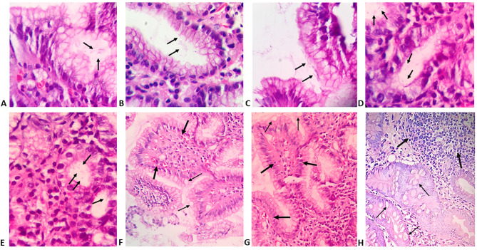
Figure 1.
Gastric biopsies specimens showing: A-C) superficial gastritis with minimal colonisation of gastric mucosal glands with few scattered H. pylori taking typical S-shaped bacilli (arrows); D,E) superficial gastritis with minimal colonisation with H. pylori taking atypical coccoid and irregular shapes (arrows) (H&E; Magnification, 400x); F,G) chronic gastritis with intestinal metaplasia and presence of papillary configuration (thick arrows) with mucous-secreting cells (thin arrows); H) H. pylori associated follicular gastritis with lymphoid follicles (thick arrows) situated deeper in the gastric mucosa and associated with intestinal metaplasia (thin arrows) (H&E; magnification 200x).

