Figure 2.
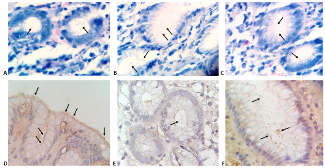
Gastric biopsies specimens showing: A-C) gastric mucosal glands with minimal colonization with H. pylori (arrows) (modified Giemsa stain; magnification 400x). D-F) H. pylori observed using immunohistochemistry staining; D) minimal colonization with typical spiral S-shaped bacilli (arrows); E) minimal colonisation with atypical coccoid forms; F) enhanced colonization with coccoid and irregular forms (IHC, magnification, 400x).

