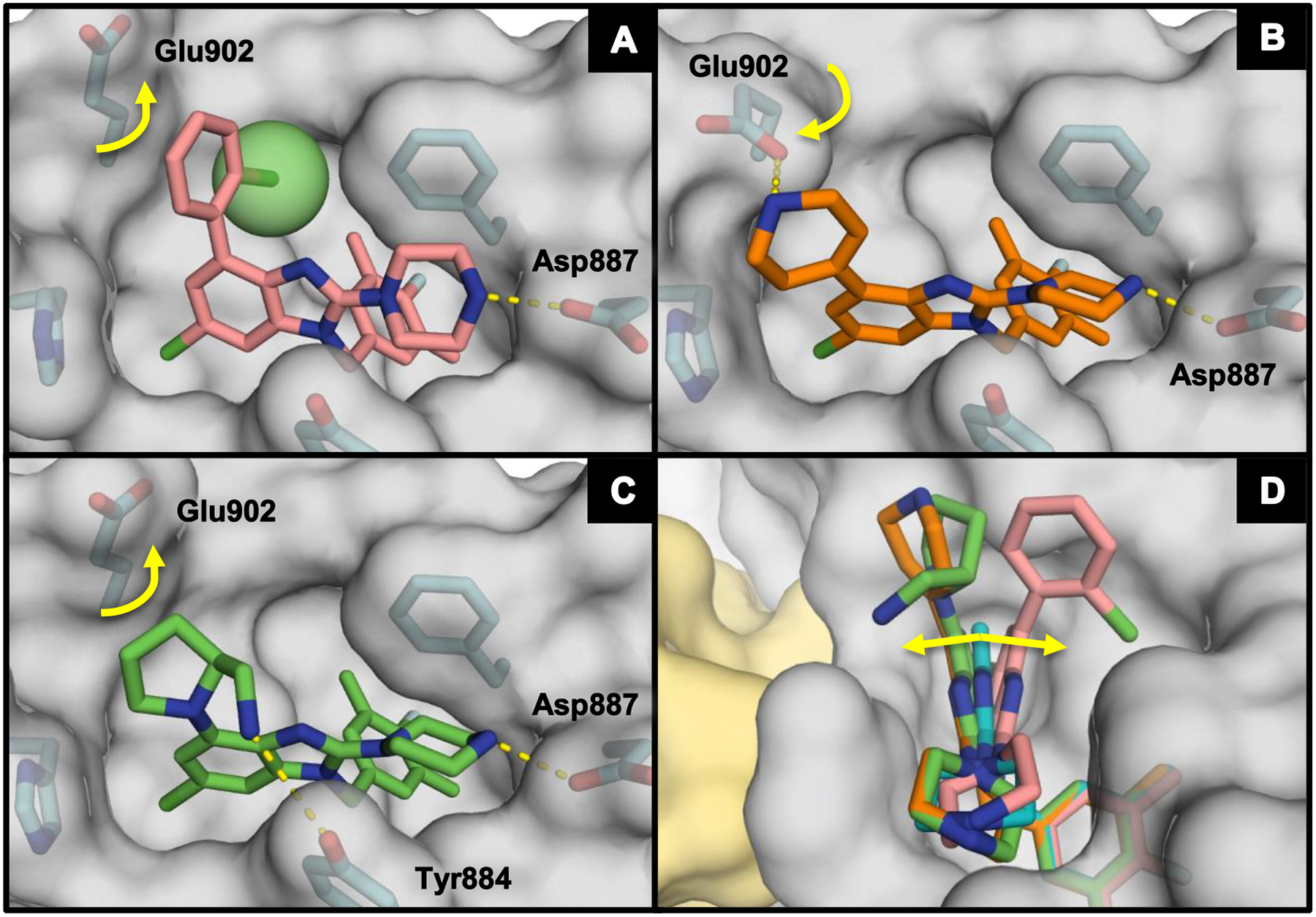
Figure 4.

X-ray co-crystal structures of (A) 38 (salmon; PDB ID code 6D5H), (B) 43 (orange; PDB ID code 6D5G), (C) 47 (green; PDB ID code 6D5E), and (D) 38, 43, and 47 overlaid with 29 (teal) bound to SOS1 in the RAS:SOS1:RAS ternary complex. The RAS and SOS1 protein surfaces are colored yellow and grey, respectively.
