Fig. 4.
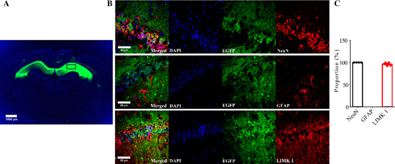
Viral expression of LIMK1-EGFP in the hippocampus. A Sample image showing EGFP fluorescent signals within the hippocampus of APP/PS1 mice 3 weeks after bilateral AAV virus injection (3-month old). Scale bar: 1000 μm. B Immunostaining images for the neuronal marker NeuN, astrocytic marker GFAP and LIMK1 showing LIMK1-EGFP signals colocalized with NeuN and LIMK1, but not with GFAP. Scale bars: 50 μm. C Summary graph showing proportion of LIMK1-EGFP expressing cells that also expressed NeuN, LIMK1 or GFAP (n = 5 sections from 5 mice)

