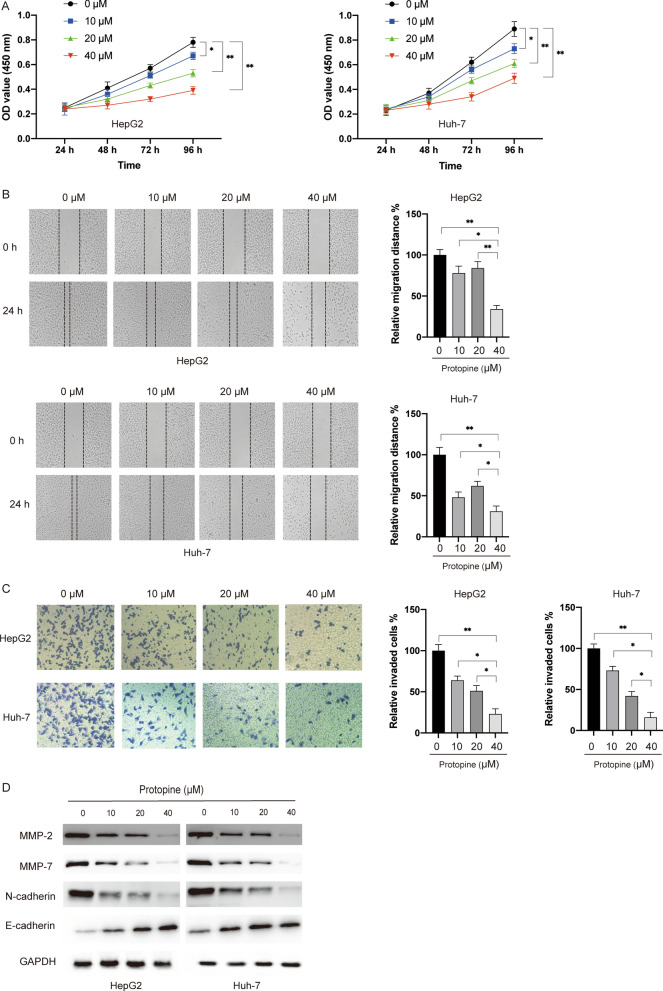
Fig. 1.
Protopine inhibited the viability, migration, invasion and EMT process of liver carcinoma cells. A Liver carcinoma cells HepG2, Huh7 and normal liver cell LO2 were treated with various doses of protopine (10 μM, 20 μM, 40 μM) for different time (24 h, 48 h, 72 h, 96 h), then cell viabilities were measured by the MTT assay. B HepG2 and Huh7 cells were treated with various doses of protopine (10 μM, 20 μM, 40 μM) for 24 h, then cellular migration was measured by the wound healing assay. C HepG2 and Huh7 cells were treated with various doses of protopine (10 μM, 20 μM, 40 μM) for 48 h, then cellular invasion was measured by the transwell assay. D HepG2 and Huh7 cells were treated with various doses of protopine (10 μM, 20 μM, 40 μM) for 24 h, then total cellular lysates were subjected to western blotting analysis with various antibodies. Data were presented as mean ± SD; *P < 0.05; **P < 0.01

