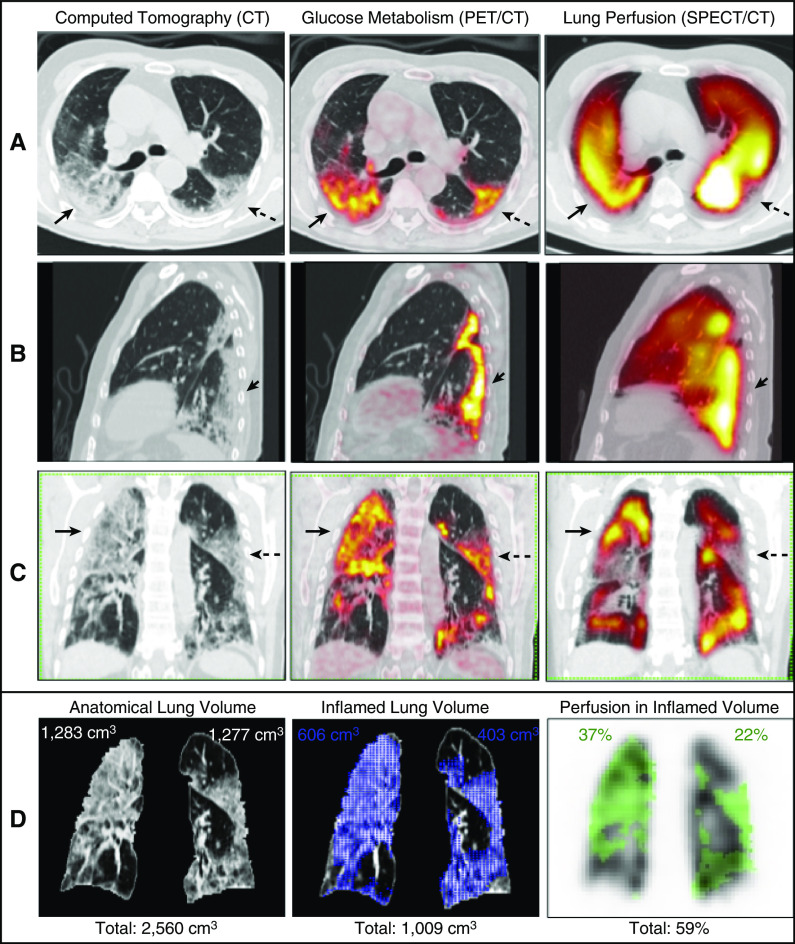
Figure 1.
(A–C) Axial (A), left sagittal (B), and coronal (C) slices from computed tomography (CT) (left), 18F-fluorodeoxyglucose (18F-FDG) positron emission tomography (PET)/CT (center), and lung perfusion single-photon-emission CT (SPECT)/CT (right). Note normal or increased lung perfusion in most hypermetabolic areas detected by PET/CT (solid arrows) compared with the apparently unaffected lung. The inflammatory abnormalities have a clear predominance in the posterior lung fields and included all consolidations or ground-glass opacities on CT images. In general, the denser the lung parenchyma on CT images, the greater the 18F-FDG uptake on PET images. Hypoperfusion (preserved vasoconstriction) is present in a few areas of 18F-FDG uptake (dashed arrows). (D) Corresponding segmented coronal slices, representative of the volumetric quantification, are shown. The segmented lungs obtained from CT images have a total volume of 2,560 cm3 (pulmonary contour in D; left). The maximal standardized uptake value of the 18F-FDG PET/CT images is 11.8. The volume of 18F-FDG uptake, representing total lung inflammation, was segmented using the mediastinal blood pool as the threshold (standardized uptake value = 1.8) and measures 1,009 cm3 (blue area in D; center). This corresponds to 39% of the total anatomic lung volume (right lung: 23%, left lung: 16%). The mask of the segmented volume of 18F-FDG uptake was transferred to the SPECT volume (green area in D; right). The counts within this mask were divided by the total counts of the SPECT image. This resulted in 59% of the total counts of lung perfusion occurring in inflamed lung tissue (right lung: 37%, left lung: 22%). Accordingly, 61% of the normal (noninflamed) lungs, which ideally should receive 100% of lung perfusion, receive only 41% of it. Therefore, this quantification estimates the total loss of hypoxic vasoconstriction in this patient. This right-to-left shunt could partially explain the severe hypoxemia of this patient. See the details of the image acquisition and quantification in the online supplement.

