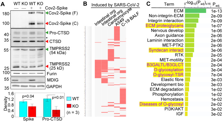
Figure 1.
Knockout of mdig diminishes the expression of genes for SARS-CoV-2 infectivity. A. Western blotting shows decreased cleavage of the transfected SARS-CoV-2 S protein and the expression of CTSD in mdig KO BEAS-2B cells. Data are representatives of at least three independent experiments. Bottom panel shows ImageJ quantifications of the cleaved SARS-CoV-2 spike protein and pro-CTSD of three independent experiments of the WT and KO cells. The data were calibrated by the density of GAPDH or histone H3. B. The down-regulated genes in the mdig KO cells as determined by RNA-seq are highly represented for those gene induced by SARS-CoV-2 in intestinal organoids (GSE149312), cardiomyocytes (GSE150392), A549 cells (GSE147507), and bronchoalveolar lavage fluid (BALF) of COVID-19 patients. C. Reactome pathway assay shows the down-regulated genes in mdig KO cells as determined by RNA-seq are mostly in the pathways of extracellular matrix (ECM) regulation and glycan metabolism (highlighted in yellow).

