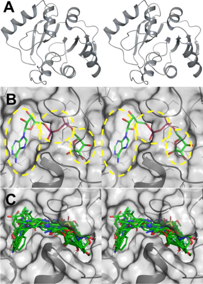
Figure 4.

ARH domain of the NSP3 protein of SARS-CoV-2. (A) Stereoview of the ribbon representation of the ARH domain, with the active site in the center. (B) Stereo view of the X-ray structure of the ARH domain depicted as a solvent-accessible surface in complex with ADP-ribose (PDB code 6W02). The adenosine, pyrophosphate, and ribose (from left to right) binding subpockets are shown by yellow broken highlights. (C) Stereoview of the superimposition of 21 top-ranking compounds docked to the NSP3 ARH domain active site (PDB code 6WCF; resolution of 1.06 Å), demonstrating sampling of all three subpockets for binding.
