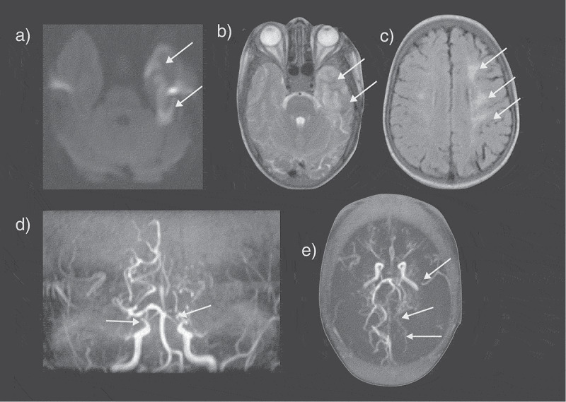
Figure 2.

An SCD patient presented with seizures and a right sided neurological deficit (B) Axial T2-weighted images shows an acute left temporal lobe infarct in the left middle cerebral artery (MCA) territory with (A) restricted diffusion on the apparent diffusion map (ADC) map (arrows). (C) The FLAIR sequence revealed extensive mature ischemic changes within the subcortical, deep and periventricular white matter of the centrum semiovale, more marked on the left (arrows). (D) MRA revealed bilateral occluded terminal internal carotid arteries (ICA) and multiple moyamoya and pial collaterals (arrows). (E) The left posterior cerebral artery (PCA) is narrowed with distal pial vessels visible (arrows). FLAIR = fluid attenuated inversion recovery
