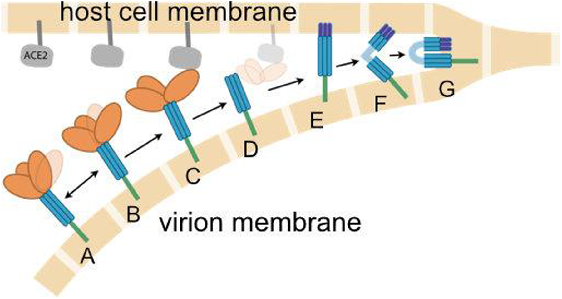
Figure 2.

Cartoon illustration of the presumed role of the spike in fusion of the viral (lower beige blocks) and host cell (upper blocks) membranes. The RBD on the S1 subunit (orange) is attached to the S2 subunit (blue), and fluctuates between (A) closed and (B) open states. When the spike approaches the ACE2 receptor (gray), the open RBD is capable of binding to ACE2 (C), leading to shedding of the S1 subunit (D), insertion of fusion peptides into the host membrane (E), additional conformational changes to co-localize the membranes (F) and eventual membrane fusion (G). Double arrows indicate reversible dynamics, while single arrows indicate presumably irreversible events. Experimental structures for states D, E and F have not been reported. Image credit: Sarina Bromberg and Carlos Simmerling
