
Figure 1. Macrophages dominate obesity-associated islet inflammation.
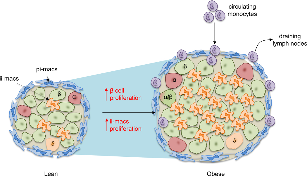
Illustrated here is a comparison of mouse islets between lean and obese conditions. In lean mice, two major populations of macrophages can be detected based on their anatomical distributions: peri-islet macrophages (pi-macs) (F4/80hi CD11c-) and intra-islet macrophages (ii-macs) (F4/80lo CD11chi). Both are islet-resident cells. In contrast, in obesity, the size of islet is increased due to increased beta cell replication and cell size. Multiple studies have demonstrated the increase of islet macrophages. However, different mechanisms have been proposed to explain obesity-associated macrophage accumulation in the islet. In one model, stressed beta cells recruit circulating monocytes which differentiate into pro-inflammatory macrophages after infiltrating into the islets. This model has been challenged by other studies showing that even though monocytes can be detected in the pancreas, they do not infiltrate into the islets. Instead, the accumulation of intra-islet macrophages is caused by local proliferation of resident macrophages.