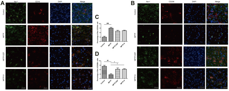
Figure 6.
Immunofluorescence double staining of inflammatory marker CD16 and anti-inflammatory marker CD206 on microglia in the substantia nigra of mice. (A) Staining of Iba-1 (green) and CD16 (classical microglia marker, red) in the SNpc and (B) quantification of the percentage of CD16+/Iba‐1+ cell. (C) Double staining of Iba-1 (microglia marker, green) and CD206 (alternative microglia marker, red) in the SNpc for immunofluorescence pictures and (D) quantification of the percentage of CD206+/Iba‐1+ cell. Scale bar is 20 µm. Data represent the means ± SEM; Statistics one-way ANOVA; ##P < 0.01, ###P < 0.001 vs Control group; *P < 0.05, vs MPTP group; n = 3.

