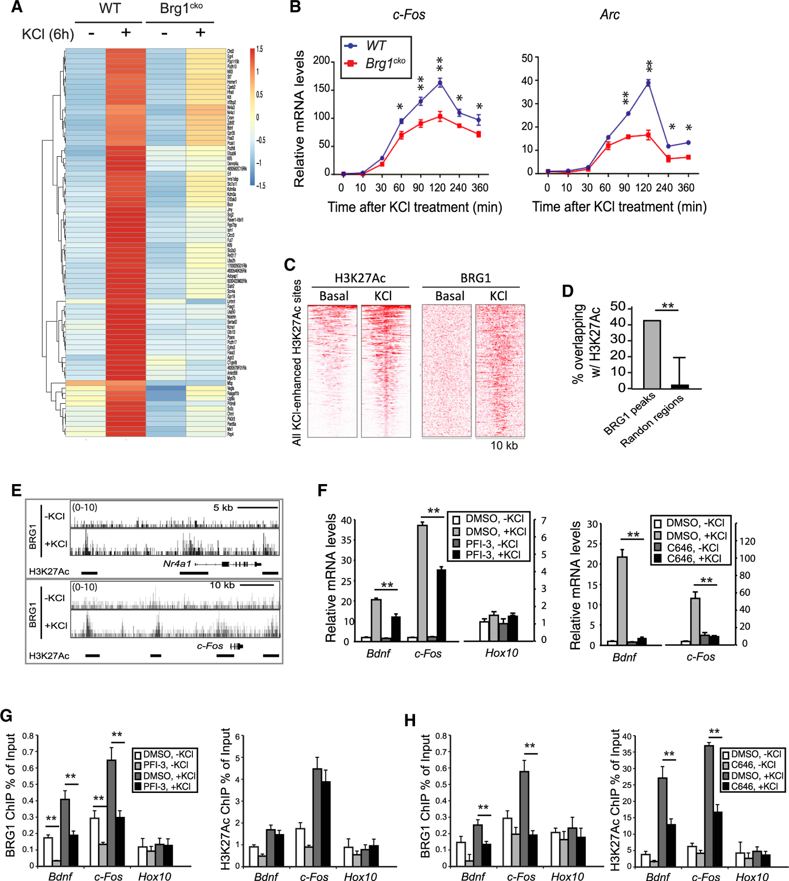
Figure 1. BRG1 regulates ARG induction and binds to H3K27Ac marked enhancers in response to neuronal activities.

(A) Heatmap showing RNA-seq signals of ARGs that were differentially expressed in wild-type and Brg1cko neurons at 6 h after KCl treatment.
(B) qRT-PCR analyses of c-Fos and Arc mRNA in cultured Brg1cko and control neurons after KCl treatment.
(C) Heatmap of H3K27Ac and BRG1 ChIP-seq signals in 10-kb regions surrounding all H3K27Ac peaks that showed increased signals upon depolarization in cultured cortical neurons (1 h).
(D) Percent overlap between BRG1 peaks and randomly selected regions with H3K27Ac peaks in depolarized neurons.
(E) BRG1 ChIP-seq signals in resting and depolarized neurons around activity-induced genes Nr4a1 and c-Fos. H3K27Ac peaks are shown as bars below the BRG1 ChIP-seq plot.
(F) qRT-PCR analyses of cultured cortical neurons (6 h, ± KCl) treated with PFI-3 or C646.
(G and H) ChIP-qPCR assays of BRG1 and H3K27Ac levels in cultured cortical neurons treated with or without PFI-3 (G) or C646 (H) (0.5 h, ± KCl).
Student’s t test (n = 3), *p < 0.05, **p < 0.01. See also Figures S1 and S2 and Table S1.
