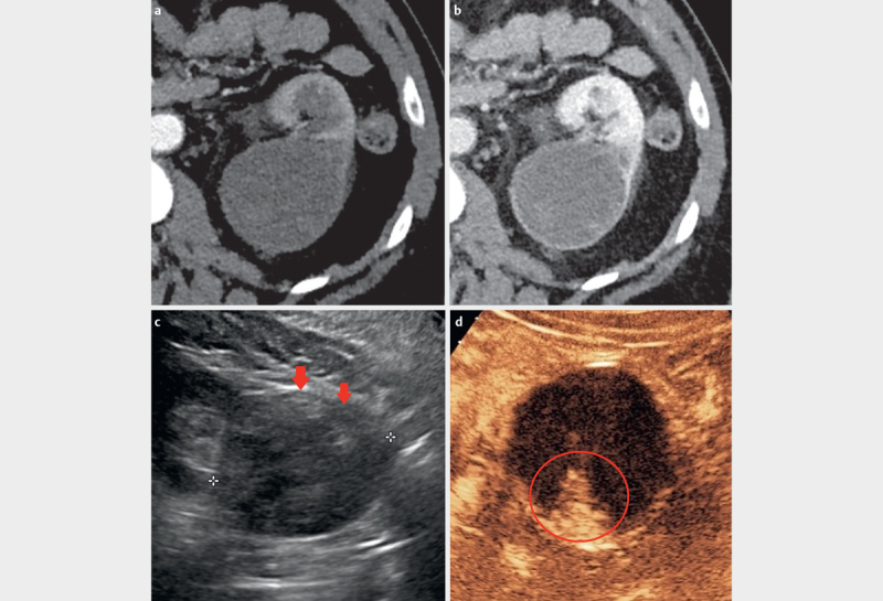
Fig. 6.

Highly suspicious lesion; Bosniak IV. a 6 cm hypodense lesion in the pars intermedia of the left kidney with a density between 20 and 30 Hounsfield units on contrast-enhanced CT (arterial phase). b In the portal venous phase, the lesion shows density values up to 40 Hounsfield units, but with enhancement of the septae. c On the B-mode image, low echoes with solid, echo-rich parts are displayed (arrows). d CEUS shows strong partial enhancement (circle) extending to the center, matching the vessels at the edges of the lesion. The lesion was classified as a partially cystic, partially solid tumor (Bosniak IV). Histologically it was a papillary renal cell carcinoma.
