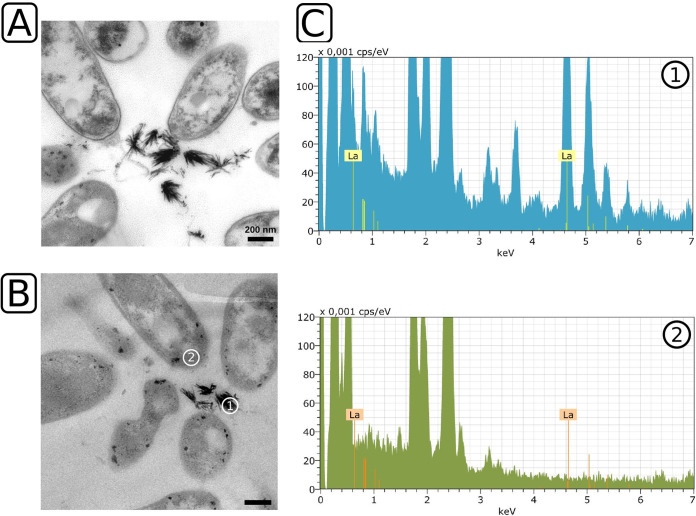
FIG 1.
Ultrathin-section transmission electron microscopy and EDX analysis of Beijerinckiaceae bacterium RH AL1 grown with 10 μM lanthanum. Strain RH AL1 was grown in MM2 medium (pH 5 to 5.5) with methanol (1% [vol/vol]) as the carbon source. Harvested biomass was fixed with glutaraldehyde (2.5% [vol/vol]), dehydrated with ethanol, and stained with uranyl acetate (2% [wt/vol]). Embedded samples were cut and ultrathin sections stained with lead nitrate. (A) Transmission electron micrographs were inspected with a Zeiss CEM 902 A electron microscope (Carl Zeiss AG, Oberkochen, Germany). (B and C) Representative sample areas for lanthanum crystals (area 1) and background signals (area 2) (B) were used for EDX analysis (C) with a Tecnai G2 electron microscope (FEI, Eindhoven, Netherlands). Lanthanum was detected by a multipoint-EDX analysis of the sample areas using an energy-dispersive X-ray spectrometer system, Quantax 200, with an XFlash detector (model 5030; Bruker, Berlin, Germany). Scale bar = 200 nm. keV, kilo electron-volt; cps/eV, counts per second per electron volt. The X-ray energy for lanthanum is highlighted (La, 4.65 keV).

