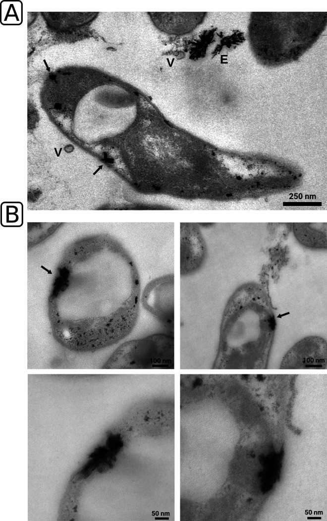
FIG 3.
Ultrathin-section transmission electron microscopy and screening for intracellular lanthanum deposits. (A and B) Lanthanum deposits were identified based on morphology and more closely inspected (B, lower panel) for proximity to the cytoplasmic and outer membrane. Black arrows indicate crystalline accumulations. E, extracellular crystalline accumulations; V, outer membrane vesicle.

