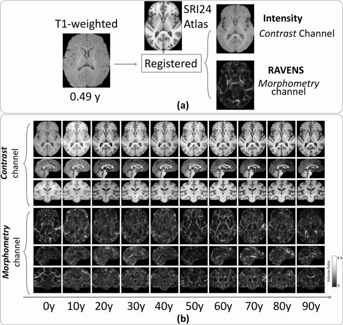
Figure 2:
Explicit split of a 3D T1w image into two 3D images representing two channels of information (contrast and morphometry). (a) A subject’s T1w image was registered to the SRI24 atlas, leading to a 3D registered intensity image (the first channel, contrast information) and a 3D RAVENS image (the second channel, morphometry information), both residing in the SRI24 atlas space. (b) Randomly-chosen subjects in every ten years of the age range for their two channels of images. Each column shows the MRI slices in the axial (top row), sagittal (middle row), and coronal (bottom row) planes. All images resided in the SRI24 atlas space.

