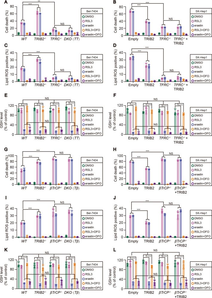
Fig. 6. TFRC and βTrCP were both required for TRIB2-desensitized ferroptosis and -reduced ferroptosis-associated lipid ROS generation.
Cell death (A, B), lipid ROS generation (C, D), and GSH levels (E, F) were measured by staining with Sytox green (A, B) and C11-BODIPY (C, D) following flow cytometry and a GSH assay kit (E, F), respectively in Bel-7404 and SK-Hep1 cells with or without knockout and overexpression of TRIB2, in the presence or absence of TFRC following treating with RSL3 (5 µM, 12 h) or erastin (10 µM, 12 h) with or without DFO (25 µM, 12 h). Cell death (G, H) and lipid ROS generation (I, J) and GSH levels (K, L) were measured by staining with Sytox green (G, H) and C11-BODIPY (I, J) following flow cytometry and a GSH assay kit (K, L) in Bel-7404 and SK-Hep1 cells with or without knockout and overexpression of TRIB2, in the presence or absence of βTrCP following treating with RSL3 (5 µM, 12 h) or erastin (10 µM, 12 h) with or without DFO (25 µM, 12 h). Data were analyzed by one-way ANOVA and expressed as mean ± SD from three independent experiments. NS non-significance; **P < 0.01; ***P < 0.001.

