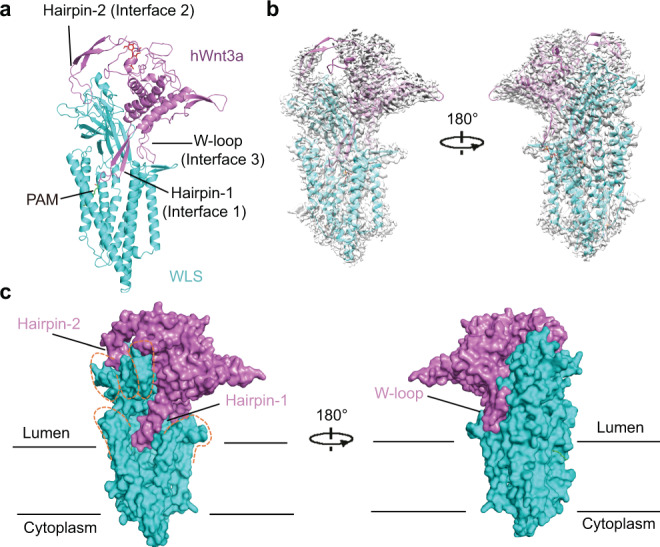
Fig. 1. Cryo-EM structure of human WLS in complex with Wnt3a.

a Overall structure of WLS-Wnt3a complex. Structure is shown in cartoon with WLS colored in cyan and Wnt3a colored in violet. The sugar moiety on Wnt3a is shown as red sticks, and PAM modified on Wnt3a is shown as green sticks. Key structural elements of Wnt3a at interfaces are indicated. b Fit of WLS-Wnt3a complex model with the 2.2 Å cryo-EM map. Wnt3a is colored in violet and WLS is colored in cyan. The map is generated from the 2.2 Å map in Chimera65 at contour level of 0.2 with dust hidden. c Complex structure is shown in surface. Model is colored in same pattern as in Fig. 1b. The contour of hand shape of WLS is shown in orange dash lines. All structural figures are generated with PyMOL66.
