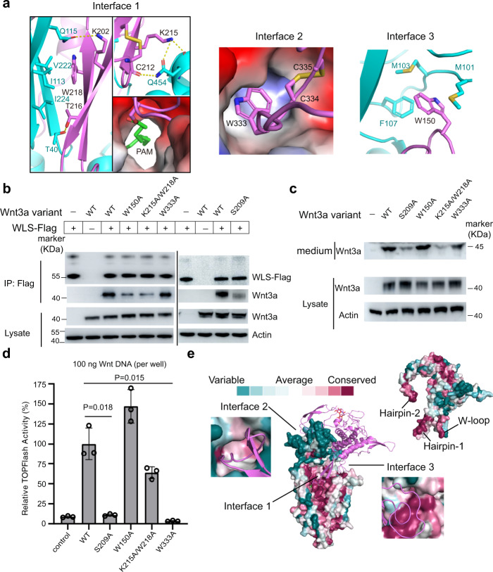
Fig. 3. Interfaces between WLS and Wnt3a.
a Three major interfaces between WLS and Wnt3a and the interaction details. Residues are indicated as one-letter codes here and in all other figures with specific residues highlighted. b Interaction validation between WLS and different Wnt3a variants through co-IP. In total 2.5 µg /well (six-well plate) of plasmids were co-transfected into Hela cells with Wnt3a: WLS DNA ratio at 1:1. Experiments were independently performed for three times with similar results. c Secretion of Wnt variants, detected by western blotting. Experiments were independently performed for three times with similar results. d Signaling activity for different Wnt3a variants. Activity was measured by TOPFlash luciferase reporter assay. Normalized activity for WT Wnt3a is taken as 100%, and activity of Wnt3a mutants is shown as percentage activity compared to WT Wnt3a. 100 ng of Wnt3a DNA per well (24-well plate) was used during transfection. All histograms were generated from n = 3 independent measurements by GraphPad Prism 9. Statistical analysis was performed by two-sided test; mean ± S.D. e Conservation surface of Wnt3a and WLS. The highly conserved regions involved in binding at interfaces are indicated.

