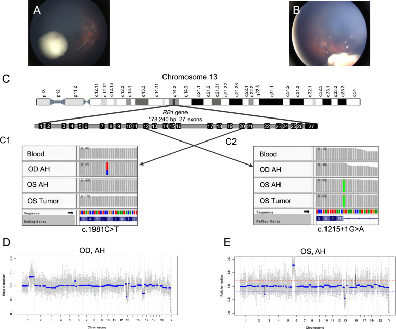
Fig. 1. Comparison of clinical images at diagnosis and genomic profiles between right and left eyes.
A Fundus photograph of the right eye shows a cohesive, creamy white endophytic mass with predominantly scattered dust-like seeds overlying the apex and a few spherical vitreous seeds at the base of the tumor, consistent with IIRC Group C retinoblastoma. B Fundus photograph of the left eye shows a creamy white endophytic mass with intratumoral vasculature, diffuse large spherical seeds, and some dust-like seeds, consistent with IIRC Group D retinoblastoma. C Integrative genomics viewer (IGV) displays the somatic RB1 pathogenic variants in the right and left eyes. The RB1 gene is located on chromosome 13 and has 178,240 base pairs with 27 exons. Here, each vertical bar represents one base pair and gray color indicates there is no change compared with the human reference genome (hg19). C1 The right eye demonstrated a missense mutation (c.1981C>T) in exon 20, seen as the red-and-blue bar found only in the OD AH sample. This represents the second hit unique to the right eye; it is not present in the blood, OS AH, or OS tumor samples. C2 The left eye demonstrated a splice donor variant mutation (c.1215+1G>A) in exon 12, seen as the matching green bars in the OS AH and the OS Tumor samples. This mutation is unique to the left eye and is only seen in OS samples; it is not present in the blood or OD AH samples. The right eye (D) and left eye (E) demonstrate non-identical somatic copy number alteration (SCNA) profiles. The right eye demonstrated 1q gain, 13q loss (germline), and 16q loss. The left eye demonstrated 6p gain and 13q loss (germline). Of note, the 6p peak amplitude seen in the right eye remains below the 20% deflection threshold (represented by the red line) to be considered a true gain.

