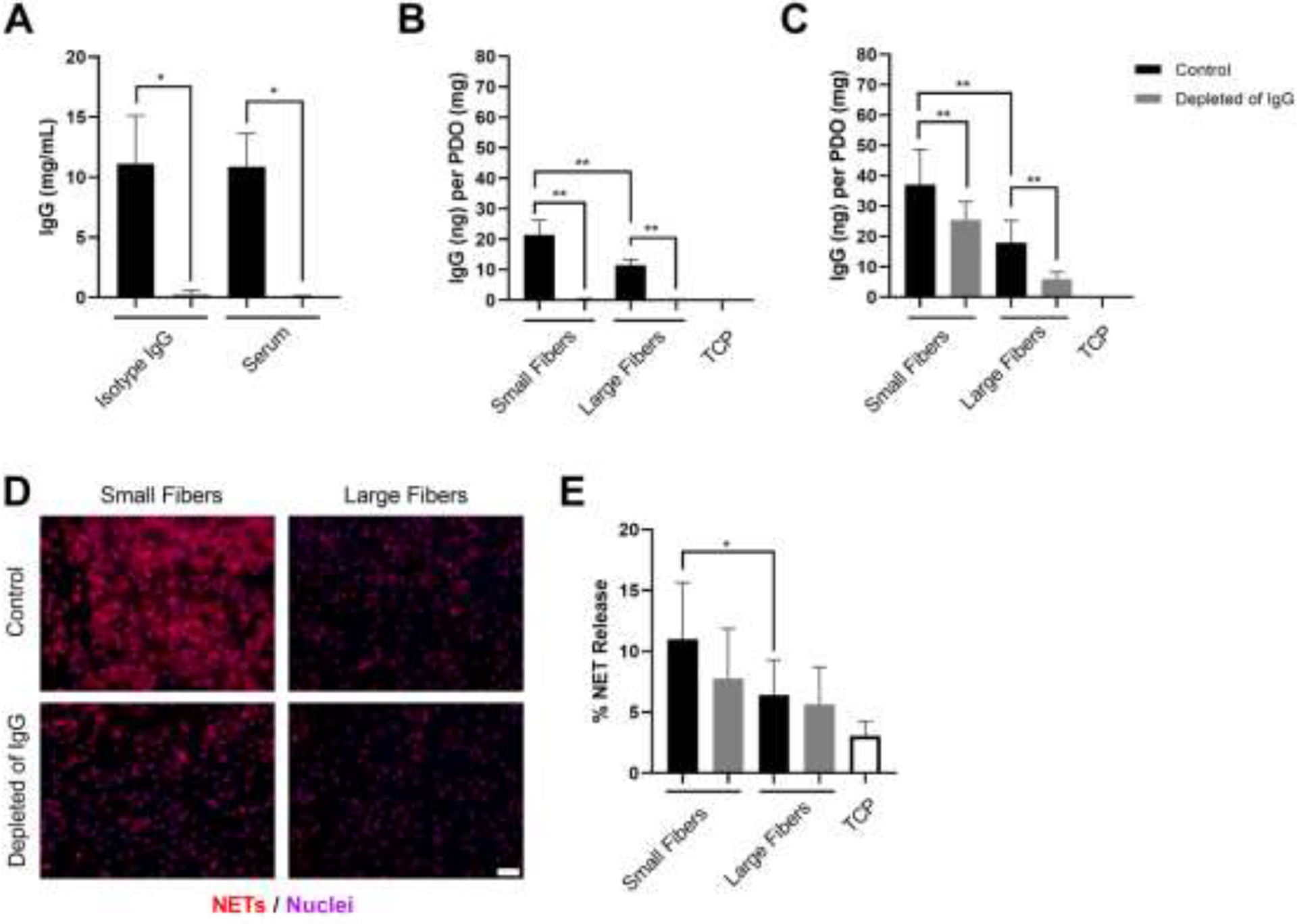
Figure 3.

Decreased IgG adsorption decreases NET release on the small but not large fiber electrospun biomaterials. (A) Concentration of IgG in isotype control (n = 3) and serum (n = 3) before and after depletion with Dynabeads™ protein G. The depletion procedure reduced the concentration of IgG by 97.4% and 99.3% for the isotype control and serum, respectively. The stock concentration of the isotype control was 11 mg/mL. * p < 0.05 was determined using a Kruskal-Wallis test with Dunn’s multiple comparisons procedure. (B) IgG adsorption on the electrospun biomaterials prior to seeding the neutrophils. (C) IgG adsorption on the electrospun biomaterials after 3-hour culture with neutrophils. The controls hydrated with cell culture media (n = 4) adsorbed significantly more IgG compared to the samples hydrated with IgG-depleted cell culture media (n = 4) both prior to neutrophil seeding and after 3-hour culture. ** p < 0.0001 was determined using an ANOVA and Holm-Sidak’s multiple comparisons test. (C) Fluorescent micrographs of neutrophils on the electrospun biomaterials at 3 hours after seeding. Staining of NETs (red) and nuclei (purple) reveals that decreasing IgG down-regulates NET release on small fibers. Scale bar is 50 μm. (D) Percent NET release at 3 hours as quantified by the ELISA for NET-disassociated MPO. The quantification of percent NET release (n = 4) indicates that decreasing IgG on the small fiber biomaterials decreases NET release. * p < 0.05 was determined using an ANOVA and Holm-Sidak’s multiple comparisons test. For all graphs, the data represent the mean ± standard deviation of three independent experiments with unique donors. Individual donor data are shown in Supplementary Figure 1. Raw data are available in Supplementary Spreadsheet 1.
