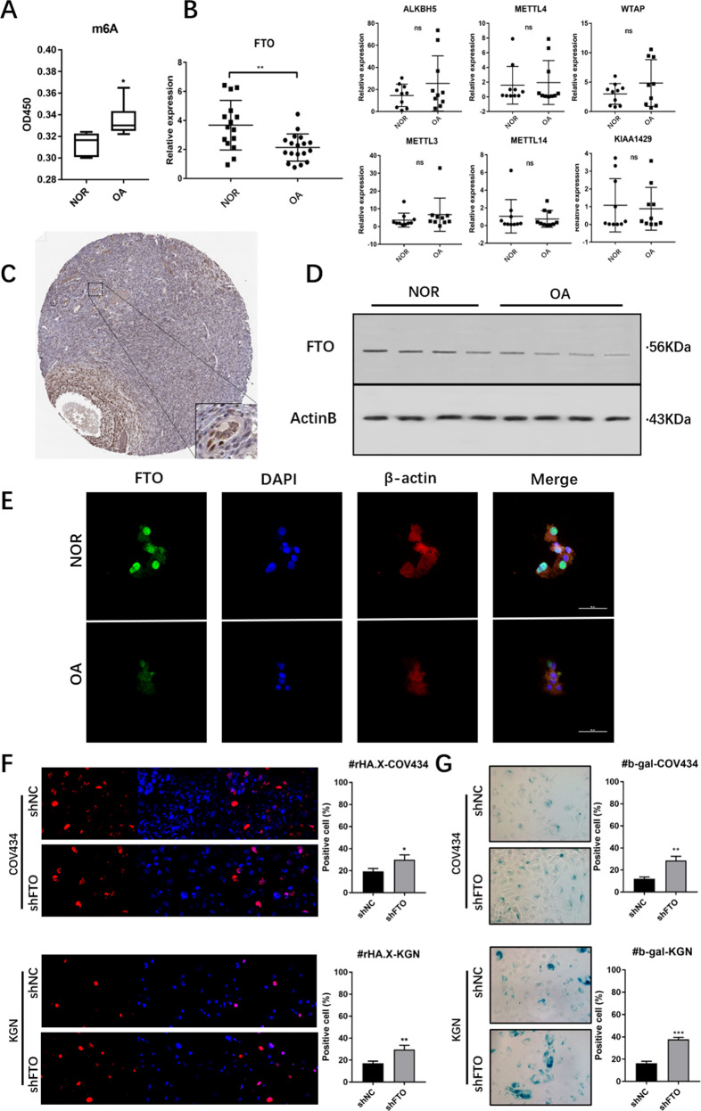
Fig. 1. Levels of RNA m6A and expression of FTO in GCs of OA patients and controls.
A Increased m6A modification in GCs of aged ovary (OA) compared to the normal ovarian reserve group (NOR), n = 6. B RT-PCR shows the mean expression levels of m6A-associated genes in the GCs in OA and NOR; for FTO, n = 15; and for other genes, n = 10. C Immunohistochemistry (from www.proteinatlas.com) showed the specific expression of FTO in granulosa cells of ovarian tissue. D, E Western blot and immunofluorescence analyses showing the downregulation of FTO in aged granulosa cells compared to young granulosa cells. F Immunofluorescence of γH2A. X and G β-galactosidase staining in FTO-silenced KGN and COV434 cells more readily enter senescence induced by hydrogen peroxide. All experiments were performed three times. The error bars indicate SD; *p < 0.05, **p < 0.01, ***p < 0.001.

