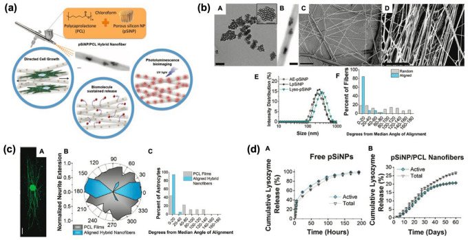
Fig. 3.
Polymer (PCL) scaffold containing pSiNPs. a Schematic illustration of pSiNPs/PCL hybrid nanofiber prepared by spray nebulization. b Transmission electron microscope (TEM) images of the hybrid formulations (A: as-prepared pSi, B: pSiNPs-embedded hybrid nanofiber, C: non-aligned hybrid nanofiber, D: aligned hybrid nanofiber) and their characterization results (E: diameter distribution of hybrid nanofibers, F: average angle of deviation of fibers from the median alignment angle of non-aligned-/aligned-nanofibers). c Directed growth study of a single astrocyte cell on the hybrid pSiNPs-PCL nanofibers, and fluorescent staining signal analysis of rat dorsal root ganglion. A: Fluorescent microscopy image of the whole dorsal root ganglia (DRG) incubated with aligned PCL nanofibers containing neurofilament (NF200)-loaded pSiNPs (incubation time: 72 h, scale bar = 500 μm). B: Normalized polar histogram of neurite growth with the aligned hybrid nanofibers (blue) and control PCL films (gray). C: the average angle of deviation of astrocyte from the median alignment angle of aligned hybrid nanofibers (blue) and control PCL films (gray). d The cumulative analysis of lysozyme amount (gray) and activity (blue) released from the nanofibers containing lysozyme-loaded pSiNPs. A: lysozyme-loaded pSiNPs without polymer hybrid, B: PCL nanofibers containing lysozyme-loaded pSiNPs. The Copyright (2018) Wiley–VCH [30]. (Color figure online)

