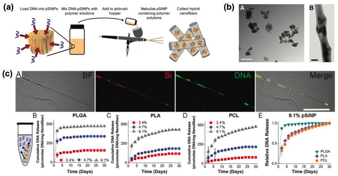
Fig. 5.
Polymer (PLGA/PLA/PCL) scaffold containing DNA-loaded pSiNPs. a Schematic illustration of the preparation of hybrid nanofiber containing DNA-loaded pSiNPs. b TEM images of A: DNA-loaded pSiNPs, B: PLGA hybrid nanofiber containing DNA-loaded pSiNPs (Scale bar – A: 200 nm, B: 2 μm). c Bright-field (BF) image and fluorescence images of PCL nanofibers. A: Si—pSiNPs image, DNA—FAM signal within DNA, Merge—the merged image of BF, Si, and DNA. Scale bar: 10 μm. B–E: Release profile of hybrid nanofiber elution in PBS at 37 °C. The supernatant was collected and changed every 48 h. B: PLGA-based nanofibers, C: PLA-based nanofibers, D: PCL-based nanofibers. Inset percent: DNA-concentration within pSiNPs. E: The comparison of the DNA release rate from the three types of nanofibers containing DNA-containing (9.1%) pSiNPs. The Copyright (2020) The Royal Society of Chemistry [33]

