
Fig. 2. Icariin mitigates glucocorticoid induced osteoporosis in vitro and in vivo.
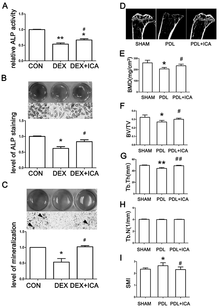
(A) (B) ALP activity and ALP staining after SaoS-2 cells being treated with same volume of DMSO (CON), 10μM DEX, and 10μM DEX plus 10−7M icariin (DEX+ICA) for 24 h. (C) ARS staining of SaoS-2 cells cultured by the osteoinductive conditioned medium containing solvent, DEX or DEX+ICA for 14 days with changes of fresh treatment medium every 2-3 days. All experiments in (A, B, C) were repeated at least three times, and the data were expressed as mean ± SD. *p < 0.05, **p <0.01 vs CON, #p < 0.05 vs DEX. (D) Representative micro-CT images of the tibia. (E) BMD assessed by micro-CT. (F)(G)(H)(I) Histogram of trabecular structure parameters of proximal tibia. *p < 0.05, **p <0.01 vs SHAM, #p < 0.05, ##p <0.01 vs PDL; n=8 for each group.