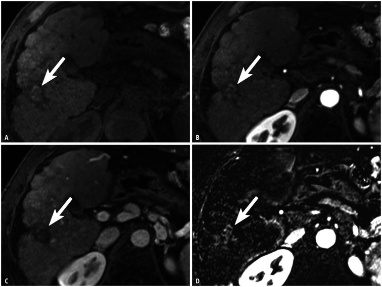
Fig. 3. HCC treated with a combination of transcatheter arterial chemoembolization and radiofrequency ablation in a 61-year-old male with cirrhosis due to chronic hepatitis B.
A. Post-treatment precontrast T1-weighted MR image shows a 2.5-cm hyperintense lesion (arrow) in hepatic segment VI. B–D. Ordinary arterial-phase image of gadoxetate acid-enhanced MRI does not depict arterial phase hyperenhancement (B), and portal venous phase image does not show washout of the lesion (arrows) (C). However, (D) arterial subtraction image shows hyperenhancement of 2.2-cm nodular enhancing area (arrow) along the posterolateral margin of the treated lesion. Therefore, the treated lesion was categorized as LR-TR nonviable based on ordinary arterial-phase images, whereas this category was changed to LR-TR viable when arterial subtraction images were used. Surgical pathology confirmed this lesion as nonviable HCC with 100% necrosis. This was a false-positive case based on the arterial subtraction images. HCC = hepatocellular carcinoma, LR-TR = Liver Imaging Reporting and Data System treatment response

