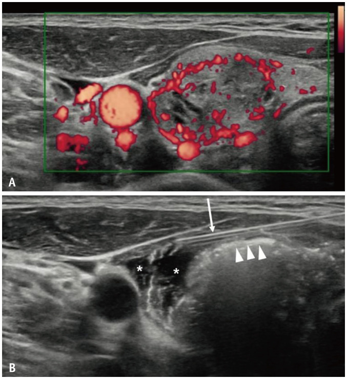
Fig. 2. Anterolateral hydrodissection and venous ablation techniques.

A. Blood vessels surrounding the nodule are clearly visible in the power Doppler ultrasonography image acquired before radiofrequency ablation. B. After completion of venous ablation, a compact filling of hot air bubbles is observed (arrowheads). A thin needle is inserted around the nodule for hydrodissection (arrow). An anechoic area was formed by the 5% dextrose injected for hydrodissection (*).
