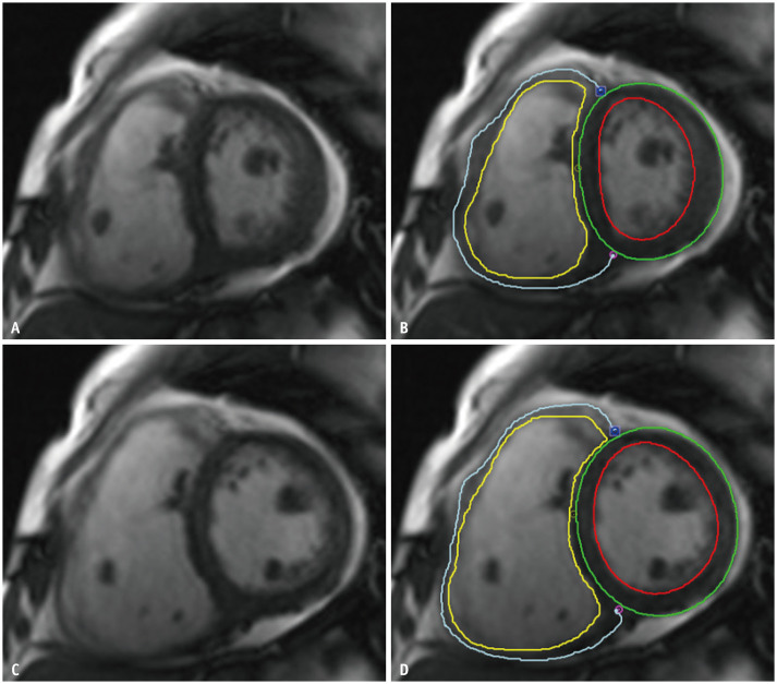
Fig. 2. Short-axis MR images with RV manual contouring during end-systole (A, B) and end-diastole phases (C, D).
Yellow and blue lines indicate endomyocardial and epimyocardial RV borders, respectively. Left ventricular endomyocardial and epimyocardial borders are drawn in red and green lines, respectively. Myocardial trabeculation and papillary muscles are excluded from the contouring. RV = right ventricle

