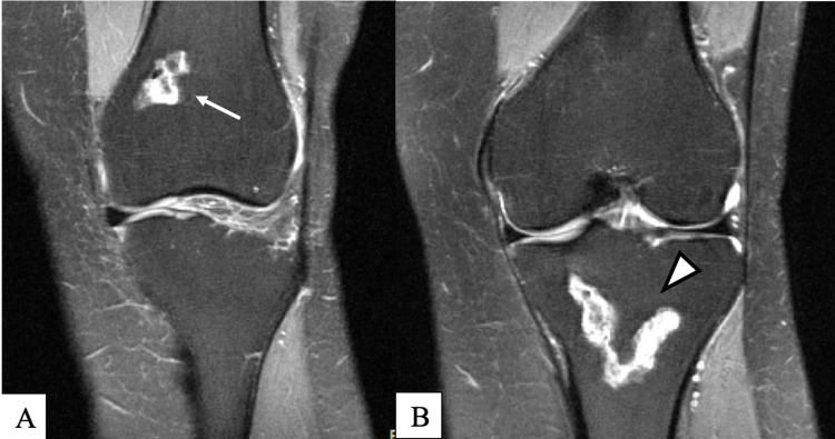
Figure 1. MRI dated June 2019.
Proton density fat saturation without contrast; coronal view demonstrates bone infarct of the distal femoral metaphysis extending to the proximal aspect of the medial femoral condyle (arrow) (A) and bone infarct of the proximal tibial metaphysis with extension into the tibial plateau and periosteal reaction along the tibial metaphysis (arrowhead) (B)
MRI: magnetic resonance imaging

