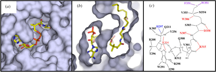
Figure 5.
The GOAT acyl-donor binding site selects for an eight-carbon acyl chain. (a) The acyl-donor binding site is exposed on the cytoplasmic face of GOAT; (b) Cutaway view of the acyl chain of octanoyl-CoA binding into a hydrophobic pocket within the enzyme internal channel. (c) Alanine mutagenesis of residues contacting octanoyl-CoA leads to either reduction (purple) or complete loss (red) of enzyme acylation activity. Figure reproduced from reference [115] under the terms of the Creative Commons CC-BY licence.

