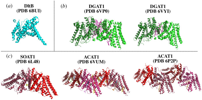
Figure 7.
Experimentally determined structures of MBOAT family members. The structure of DltB (PDB 6BUI) was determined by X-ray crystallography, with the other MBOAT structures solved by cryoelectron microscopy (DGAT1: PDB 6VPO [121], PDB 6VYI [122]; ACAT1/SOAT1: PDB 6L48 [123], PDB 6VUM [124] and PDB 6P2P [125]). Cryo-EM structures of DGAT1 solved by two laboratories report similar dimeric structures. ACAT1/SOAT1 was solved by three different laboratories, with two groups reporting tetrameric structures and the remaining group yielding a ACAT1/SOAT1 dimer. (a) DltB is shown in cyan. (b) DGAT, with each monomer in the dimer denoted by shades of green. Lipids are shown in magenta. (c) ACAT/SOAT, with each monomer depicted in shades of red. O-succinylbenzoyl-N-CoenzymeA, orange; cholesterol, yellow; nevanimibe, purple and blue; coenzyme A, violet; lipids, green. Figure created with PyMol.

