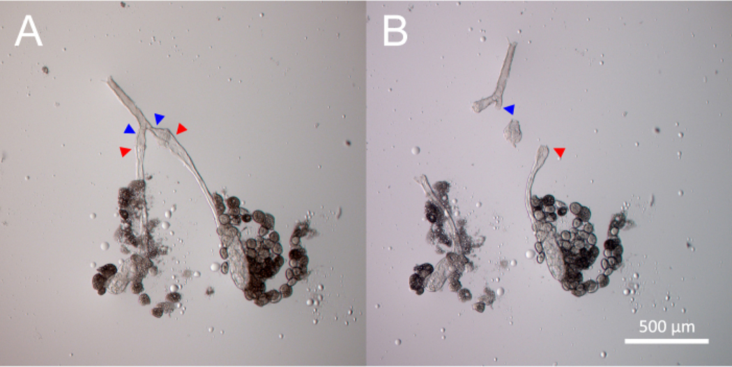
Figure 3: Dissection of the primary salivary gland ImR tumor.
The process of dissecting and isolating two primary salivary gland ImR tumors is demonstrated chronologically from Panel (A) to Panel (B), using two separate incisions. Panel (A) shows the salivary gland before tumor dissection. The red arrowheads indicate the first incision points. The blue arrowheads indicate the second incision points. The tumor lies between the red and blue arrowheads. Panel (B) shows the isolated ImR tumor after the two incisions are made to separate it from normal salivary gland tissue.

