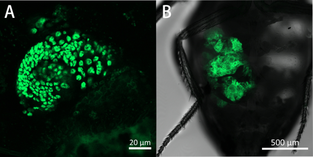
Figure 5: A G1 and G6 ImR tumor seen in WT Drosophila host abdomen on day 10 post-allotransplantation.
These are ventral views of the fly abdomen with the transplanted tumors in green. Panel (A) shows a G1 tumor on day 10 post-allotransplantation expressing eGFP (488 nm). Panel (A) is captured using a confocal microscope using a 20x lens with 0.8 NA, and 3x zoom. Panel (B) shows a G6 tumor on day 10 post-allotransplantation expressing eGFP (488 nm). Panel (B) is captured using a confocal microscope using a 5x lens with 0.25 NA, and 1x scan zoom.

