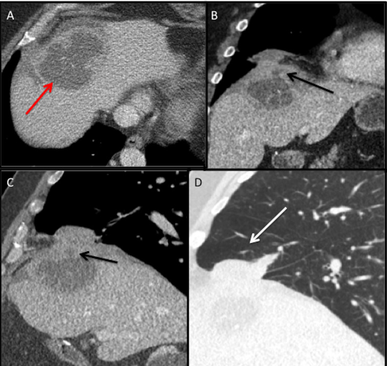
Figure 2.
CT (2019) of the chest and abdomen axial (A) section demonstrates a large liver abscess (red arrow) with a spilled gallstone in the hepatic dome. Coronal (B) and sagittal soft tissue window (C) and sagittal lung window (D) reformats demonstrate the liver abscess extending into the subphrenic space with fistulous tract through the diaphragm (black arrow) with associated right middle lobe pneumonia and abscess (white arrow).

