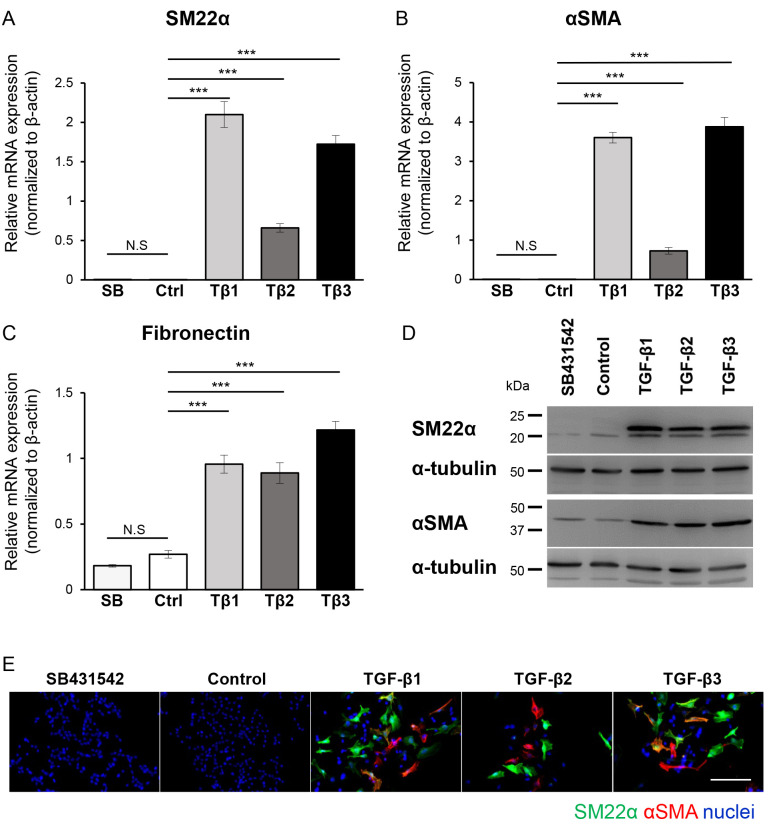
Figure 3.
EMT program in B16 melanoma cells is induced by all TGF-β isoforms. B16 cells were cultured in the absence (Ctrl or Control) or presence of TGF-β1 (Tβ1), TGF-β2 (Tβ2), or TGF-β3 (Tβ3) (3 ng/ml) or the TβRI kinase inhibitor, SB431542 (SB; 10 µM) for 72 h, followed by (A-C) RT-qPCR, (D) immunoblotting and (E) immunocytochemistry. Experiments were performed in triplicate and repeated twice. (A-C) The expression of mesenchymal markers (A) SM22α, (B) αSMA and (C) fibronectin were evaluated by RT-qPCR analyses. All RT-qPCR data were normalized to the β-actin expression. (D) The immunoblotting analysis with antibodies specific to SM22α, αSMA and α-tubulin (loading control). (E) Representative immunofluorescence images revealing staining of SM22α (green), αSMA (red) and nuclei (blue). Scale bar, 100 µm. Error bars, SD. ***P<0.001. EMT, epithelial-mesenchymal transition; TGF-β, transforming growth factor-β; TβRI, TGF-β type I receptor; RT-qPCR, reverse transcription-quantitative PCR; NS, not significant; SM22α, smooth muscle protein 22α; αSMA, α-smooth muscle actin.

