Fig. 1.
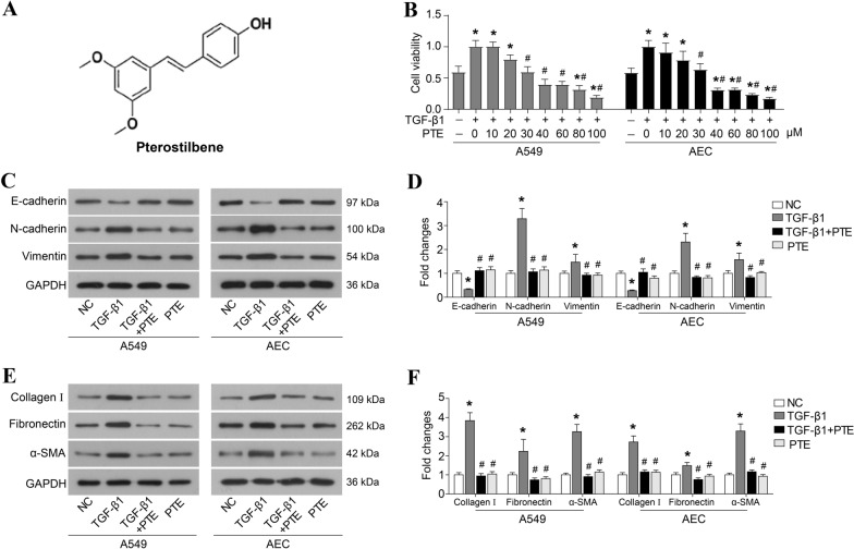
PTE inhibits TGF-β1-induced cell proliferation, EMT, and ECM accumulation. A Chemical structure of PTE. B A549 and AECs were treated with 10 ng/ml TGF-β1 and various concentrations of PTE (0, 10, 20, 30, 40, 60, 80, and 100 μmol/L) for 24 h, and cell viability was detected by CCK8 assay. A549 and AECs were treated with 10 ng/ml TGF-β1 and 30 μmol/L PTE for 24 h. The key protein of EMT (C, D), α-SMA, and ECM (E, F) were detected by western blot. All experiments were repeated three times independently, and Student’s t test or ANOVA was used to compare the differences between groups. *P < 0.05 compared with the NC group; #P < 0.05 compared with the TGF-β1 group

