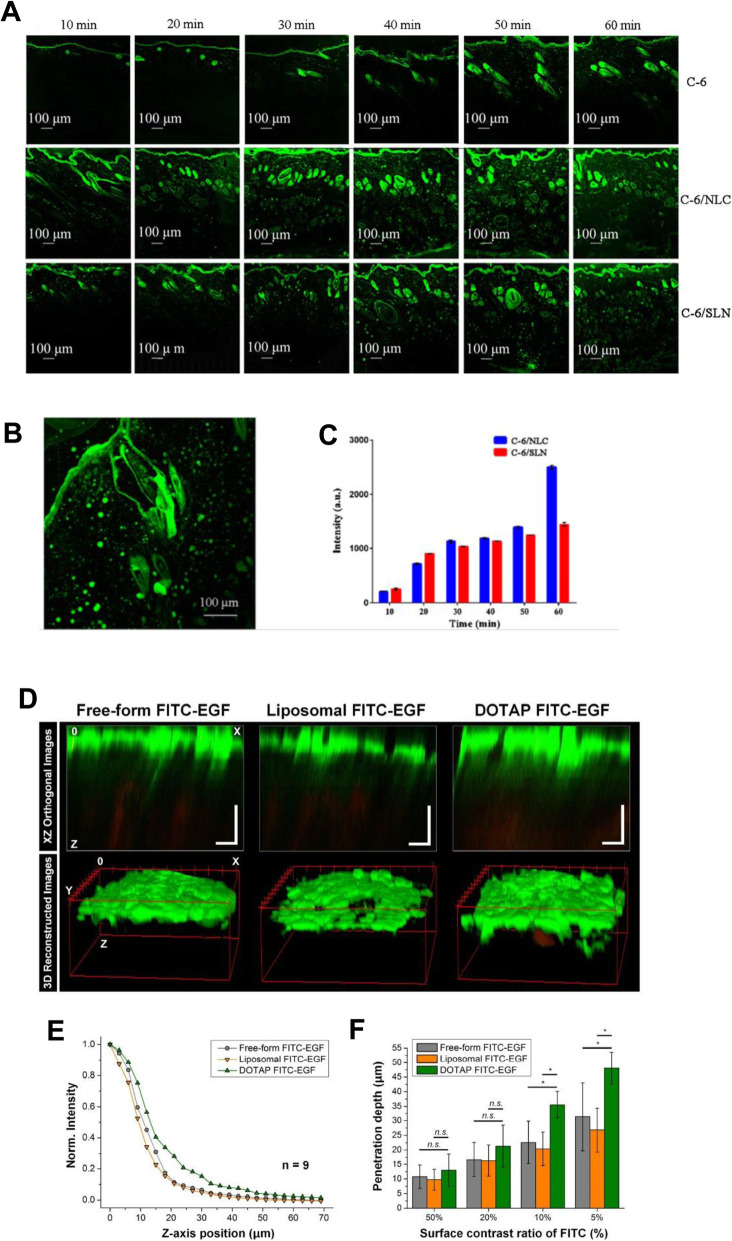
Fig. 6.
A CLSM images (100× magnification) of skin samples treated with free C-6, C-6/NLC, and C-6/SLN. B Enlarged CLSM Fig. (200× magnification). C Fluorescence intensity in receptor fluid at various times. D Reconstructed two-photon images in XZ orthogonal and 3D views. E Averaged normalized FITC-EGF signal intensity along the z-axis from the surface to the dermal layer of human skin samples. F Penetration depth of FITC-EGF with different thresholds of fluorescence intensity (50, 20, 10, and 5%) measured at the skin surface. A, B, C Reproduced from [88], copyright permission by Springer Nature 2018. D, E, F Reproduced from [89], copyright permission by OSA Publishing 2018

