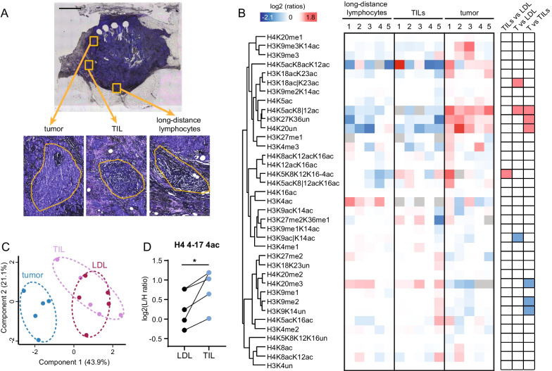
Fig. 5.
Histone PTM quantification from breast tumor samples and lymphocytes. a Crystal violet staining of a representative breast cancer section. Tumor cells (T), tumor-infiltrating lymphocytes (TILs) and lymphocytes outside of the tumor region [long-distance lymphocytes (LDLs)] were collected by LMD in five patients and analyzed by MS. Scale bar: 2 mm. b Heatmap display of the log2 of ratios obtained for the indicated histone PTMs in tumor cells, TILs and LDL in the samples described in (a). The L/H ratios of relative abundances obtained with the super-SILAC strategy (light channel: laser microdissected sample, heavy channel: spike-in standard) normalized over the average value across all samples are shown. The grey color indicates peptides that were not quantified. Right panel: modified peptides were compared by repeated measures ANOVA, followed by Tukey’s multiple comparison test. The red color indicates an increase, the blue color a decrease (p < 0.05). c PCA analysis based on histone PTM data obtained from tissue areas highlighted in (a). d Plot showing the levels of the tetra-acetylated form the H4 4–47 peptide in TILs and LDLs. Significance was assessed as in (b)

