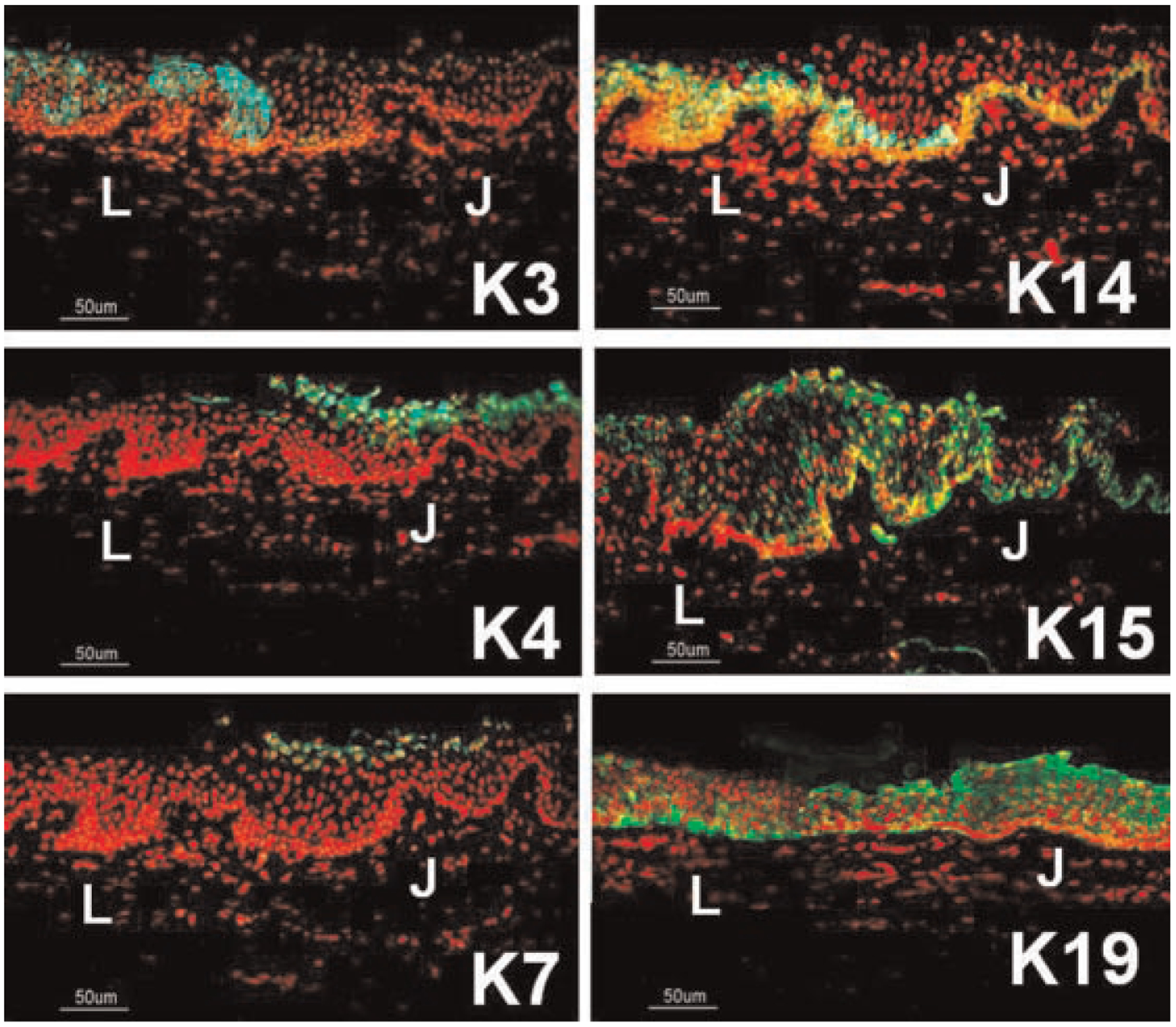
Fig. 2.

Representative images showing the immunofluorescent staining of cytokeratins (CKs) (green) in the epithelia at the limbus–bulbar conjunctiva transition zone. PI was used as nuclear counterstaining (red). CK3 was localized to the superficial limbal epithelial cells (L). CK4 and CK7 were only expressed by the superficial layers of bulbar conjunctival epithelium (J). CK14 and CK15 was confined to the basal layer of bulbar conjunctival epithelium (J), but was expressed by all layers of limbal epithelia (L). CK19 was strongly expressed by all layers of bulbar conjunctival epithelia (J), but confined to the basal limbal cells (L). Scale bar = 50 μm.
