
Figure 1. scRNA-seq of mouse SVF reveals distinct preadipocyte and mesothelial populations that express Wt1.
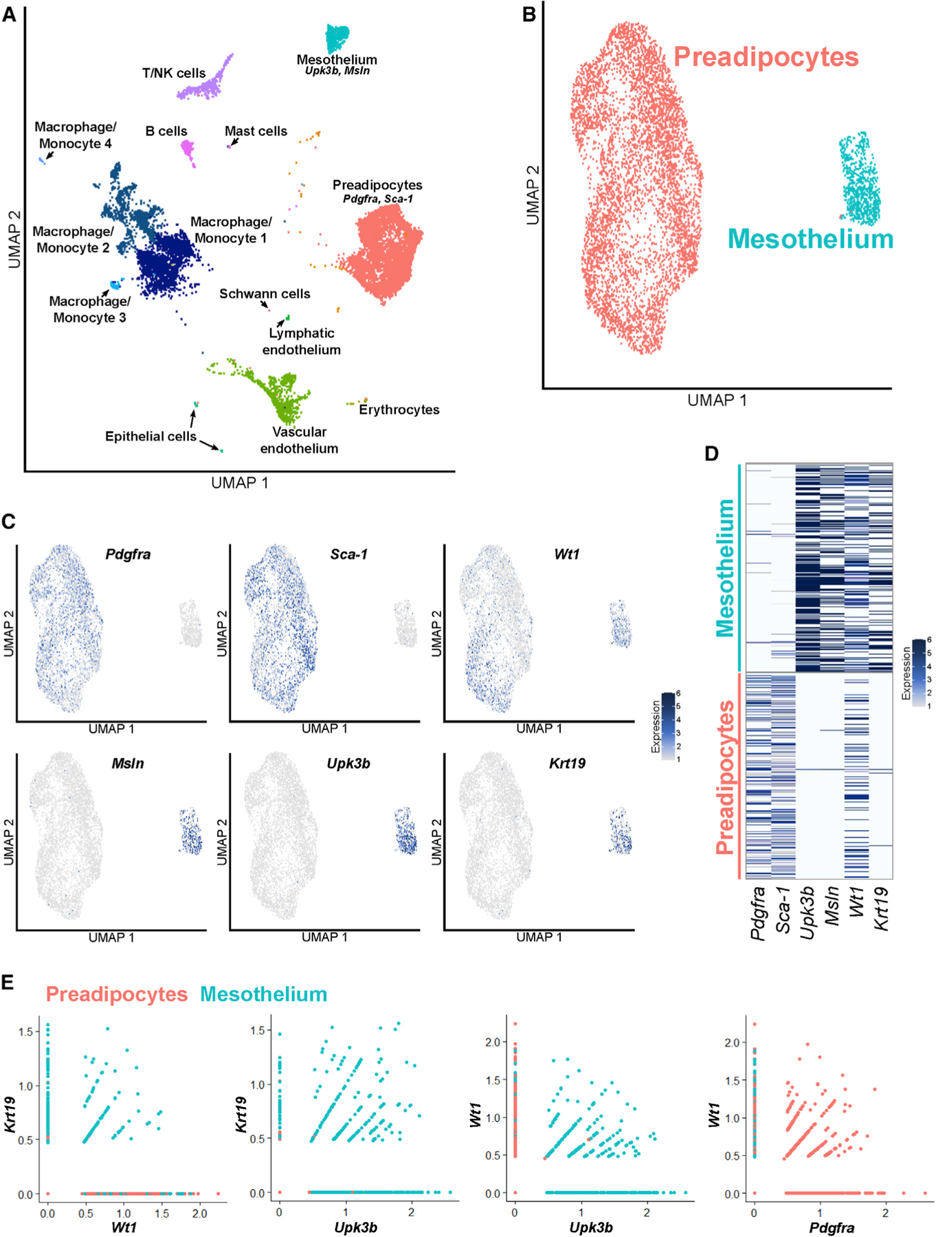
(A) Uniform Manifold Approximation and Projection (UMAP) plot of scRNA-seq performed on the SVF of mouse epididymal fat (eWAT), which identifies all expected populations, including distinct populations of preadipocytes and mesothelial cells.
(B) UMAP plot of the preadipocyte and mesothelial populations.
(C) The preadipocyte population is characterized by expression of Pdgfra and Sca-1, whereas the mesothelium expresses Upk3b and Msln. Although Krt19 expression mirrors that of mesothelial markers, Wt1 is expressed in preadipocytes and mesothelial cells.
(D) Heatmap demonstrates that Wt1 is expressed in cells expressing preadipocyte and mesothelial genes.
(E) Most cells expressing Wt1 but not Krt19 are preadipocytes, whereas virtually all Krt19-expressing cells are mesothelial. A significant subset of Wt1+ cells co-expresses Pdgfra.