
Figure 2. Wt1 is not a specific marker for the mesothelium and also labels Pdgfra+ preadipocytes.
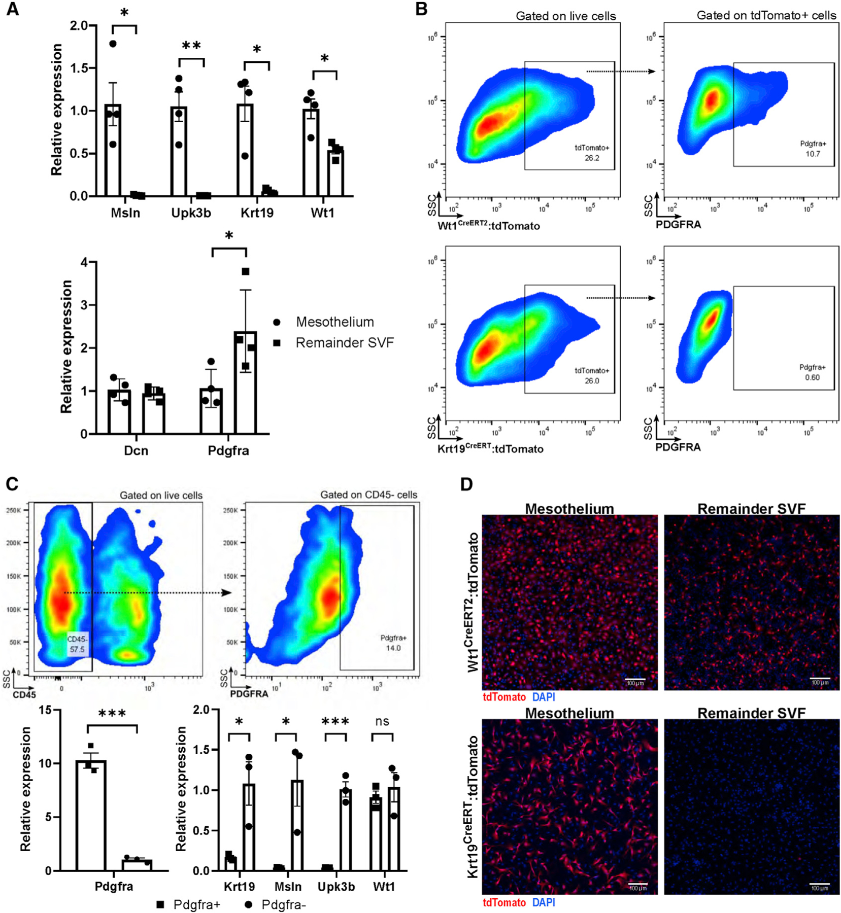
(A) The mesothelium can be digested away from the remainder of the adipose parenchyma with trypsin, a technique that highly enriches for mesothelial markers but not for Wt1, which is also highly expressed in the remainder SVF. The remainder SVF highly expresses Pdgfra, whereas Dcn is expressed by mesothelium and other stromal cells. n = 4 biological replicates of 2 mice each; mean ± SEM; *p < 0.05, **p < 0.01.
(B) Using Wt1CreERT2 and Krt19CreERT models crossed with a tdTomato reporter, flow cytometry reveals that Wt1-expressing cells contain a PDGFRA+ preadipocyte population whereas Krt19-expressing cells do not. Results are representative of two independent experiments.
(C) SVF cells isolated from wild-type mice were gated against CD45 and sorted into PDGFRA+ and PDGFRA− populations; mesothelial gene expression was restricted to PDGFRA− populations, whereas Wt1 was also present in the PDGFRA+ preadipocyte population. n = 3 groups of 2 mice each; mean ± SEM; ns, p R 0.05; *p < 0.05; ***p < 0.001.
(D) Trypsin-digested mesothelium contains a high proportion of tdTomato+ cells in Krt19CreERT and Wt1CreERT2 models, whereas the remainder SVF contains tdTomato+ cells in only Wt1CreERT2 mice. SSC, side scatter. Images are representative of 10 low-powered fields of cells cultured from 4 separate mice.