
Figure 3. Krt19-expressing mesothelial cells do not differentiate into adipocytes in vitro.
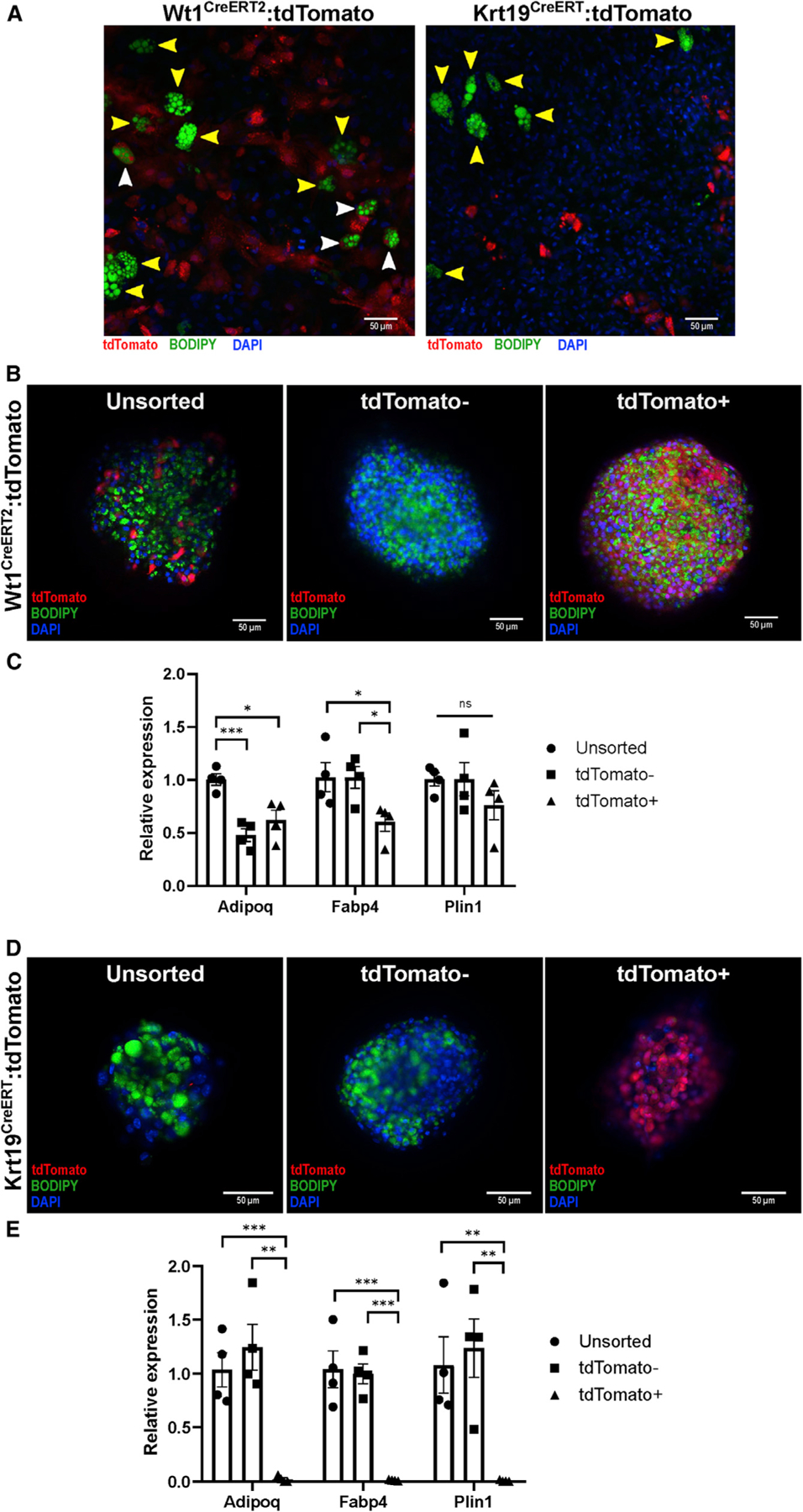
(A) SVF from Wt1CreERT2 and Krt19CreERT mice was isolated and underwent adipogenic differentiation on standard collagen-coated plates. Although adipocytes differentiated from Wt1CreERT2:tdTomato SVF contained tdTomato+ (white arrowheads) and tdTomato− (yellow arrowheads), no tdTomato+ adipocytes were found in the Wt1CreERT2:tdTomato samples. Representative images from 36 low-powered fields are shown.
(B and C) SVF was isolated from Wt1CreERT2:tdTomato mice after tamoxifen labeling, sorted into tdTomato+ and tdTomato− groups, and plated on low-adhesion plates, forming microspheres. After adipogenic differentiation, unsorted, tdTomato+, and tdTomato− cells derived from Wt1CreERT2:tdTomato mice, as demonstrated by BODIPY labeling of lipid droplet formation and qPCR for the adipocyte markers Adipoq, Fabp4, and Plin1. n = 4 microspheres derived from 3 separate mice; mean ± SEM; ns, p R 0.05; *p < 0.05; ***p < 0.001.
(D and E) SVF isolated from Krt19CreERT:tdTomato mice was sorted into tdTomato+ and tdTomato− populations, and although unsorted and tdTomato− cells differentiated normally as shown by BODIPY staining and qPCR, tdTomato+ cells did not undergo adipogenic differentiation. n = 4 microspheres derived from 5 separate mice; mean ± SEM; **p < 0.01, ***p < 0.001.