
Figure 4. Krt19-lineage mesothelial cells do not differentiate into adipocytes in vivo.
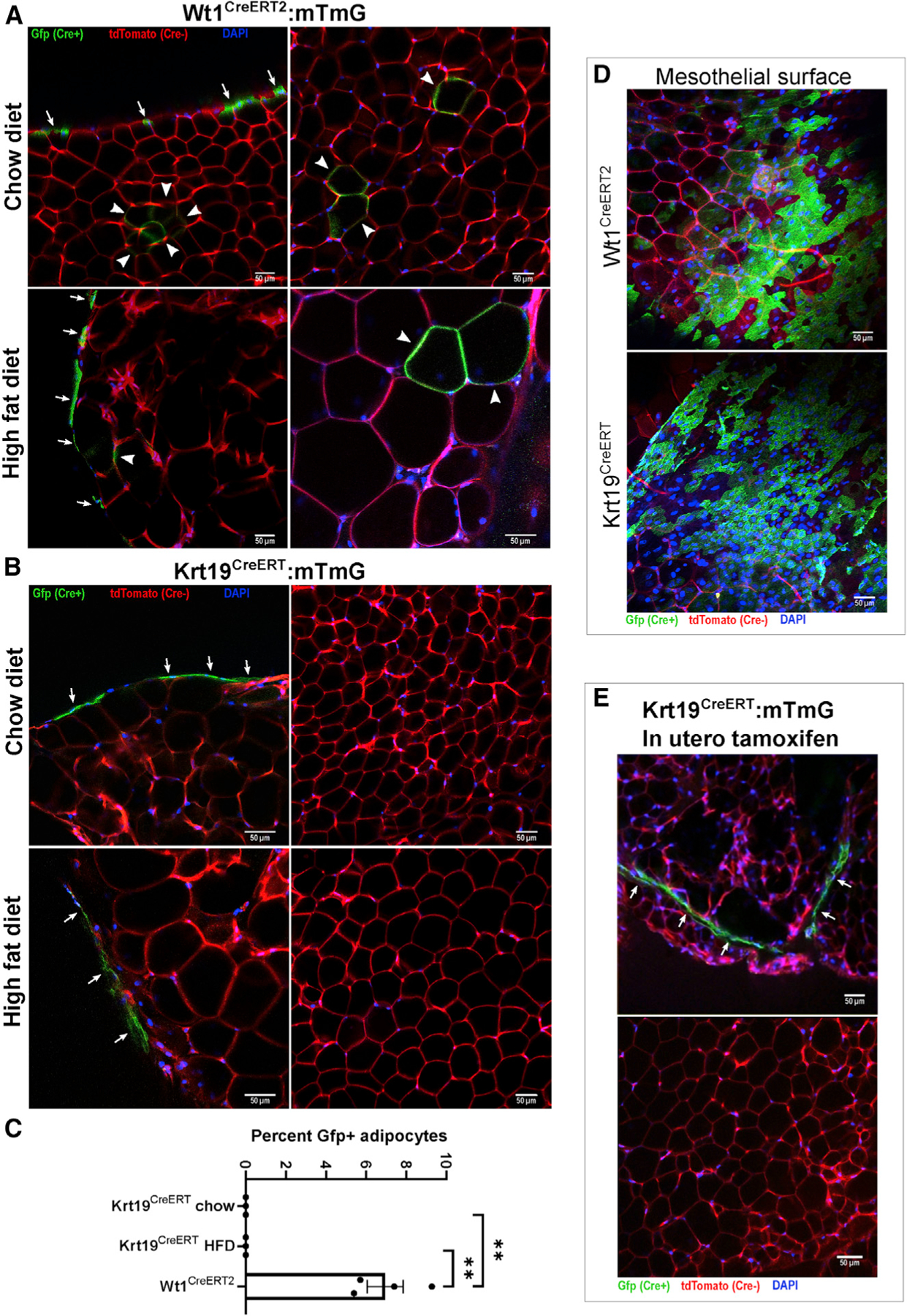
Wt1CreERT2:mTmG and Krt19CreERT:mTmG mice were injected with tamoxifen at 6 weeks of age and sacrificed at 15 weeks of age; mice receiving a high-fat diet did so starting at 7 weeks age. Whole mounts of perigonadal fat were imaged by confocal microscopy.
(A) In Wt1CreERT2:mTmG mice, labeling of the mesothelial layer (arrows) as well as multiple Gfp+ adipocytes (arrowheads) were readily observed. Shown are representative images from 26 low-powered fields obtained from 4 separate mice.
(B) In contrast, Krt19CreERT:mTmG mice only had labeling of the mesothelial layer, and no GFP+ adipocytes were observed. Representative images from 50 low-powered fields were obtained from 6 separate mice.
(C) The percentage of Gfp+ adipocytes was quantified. In the chow and HFD Krt19-CreER groups, over 2,200 and 1,600 adipocytes, respectively, were counted without any Gfp+ adipocytes observed. n = 3–4 mice, 6–10 fields per mouse; mean ± SEM; **p < 0.01.
(D) GFP+ mesothelium was readily identifiable in Wt1CreERT2:mTmG and Krt19CreERT:mTmG models.
(E) To evaluate the possibility that mesothelial cells, which develop in utero, may transition to a preadipocyte identity later in development, Krt19CreERT:mTmG dams were injected with tamoxifen at gestational day E14.5, and offspring were sacrificed at 6 weeks of age. The mesothelium contained GFP+ cells, but no GFP+ adipocytes were identified. Shown are representative images from 15 fields obtained from 2 separate mice.