. 2021 Jun 25;12(29):10054–10062. doi: 10.1039/d1sc02259h

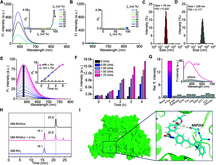
Fig. 4. Evaluation of QM-NHαfuc as an α-fuc-responsive AIE fluorescence probe. Emission spectra (λex: 543 nm) of (A) QM-NH2 (10 μM) and (B) QM-NHαfuc (10 μM) in a mixture of THF with different aqueous fractions (fw). Inset: plots of relative fluorescence intensity at 586 nm against fw. DLS data for (C) QM-NH2 (5 μM) and (D) QM-NHαfuc (5 μM) in pure water. (E) Time-dependent emission spectra (λex: 543 nm) of QM-NHαfuc (20 μM) recorded in the presence of α-fuc (2 U mL−1). Inset: plots of the relative intensity at 586 nm as a function of time. (F) Fluorescence intensity of QM-NHαfuc in the presence of different concentration of α-fuc recorded as a function of time. (G) Fluorescence response of QM-NHαfuc (20 μM) after 6 h incubation with α-fuc or other analytes. (H) HPLC monitoring of the hydrolysis of QM-NHαfuc seen in the presence of α-fuc. (I) Proposed binding mode for QM-NHαfuc within the active site of α-fuc.

Fig. 4