FIGURE 4.
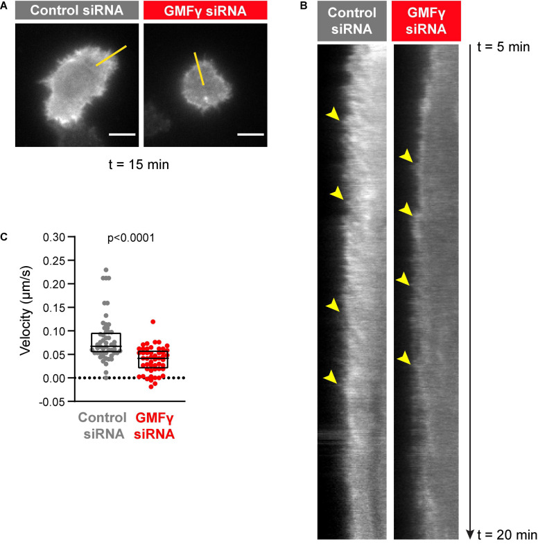
Depleting GMFγ reduces the velocity of actin retrograde flow. Raji D.3 B cells that had been co-transfected with F-tractin-GFP cDNA and either control siRNA or GMFγ siRNA were added to anti-human IgM-coated coverslips and allowed to spread for 5 min. The cells were then imaged by TIRF microscopy at 2 s intervals for 15 min (t = 5 min to t = 20 min). Video recordings of representative cells are shown in Supplementary Movie 1 (control siRNA) and Supplementary Movie 2 (GMFγ siRNA). (A) Still images of the control siRNA- and GMFγ siRNA-transfected cells shown in Supplementary Movies 1, 2 at t = 15 min. Scale bars: 5 μm. (B) Kymographs were generated along the yellow lines in (A). Arrowheads indicate the starting points of representative actin tracks for which centripetal velocities were determined. (C) The centripetal velocity (Δx/Δt) was calculated for individual actin tracks on the kymographs. Each dot on the graph is an individual actin track. The velocity was determined for 52 tracks from 9 cells (control siRNA) or 51 tracks from 11 cells (GMFγ siRNA). The Mann-Whitney U-test was used to calculate the p-value.

