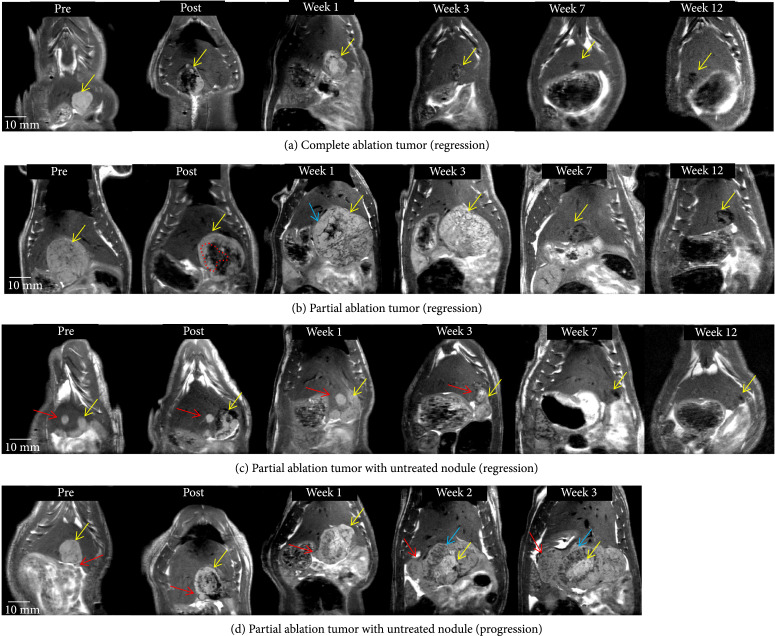
Figure 2.
Tumor response to histotripsy on T2-weighted MRI. The size and appearance of tumor (yellow arrow) in response to complete and partial histotripsy ablation are observed at different timepoints. (a) Complete ablation—pre-treatment (mildly hyperintense), posttreatment ablation zone (hypointense), and ablation zone at week 1 (mildly hyperintense), week 3 (mildly hypointense, size regression), week 7 (hypointense, size regression), and week 12 (<5 mm hypointense region). (b) Partial ablation—pretreatment (mildly hyperintense), posttreatment ablation zone (hypointense, shown by red dashed lines) and untreated tumor region (mildly hyperintense), and lesion at week 1 (mildly hyperintense) with pseudoprogression characteristics (blue arrow), week 3 (mild hyperintense, size regression), week 7 (hypointense, size regression), and week 12 (~5 mm hypointense region). (c) Partial ablation with a separate untreated nodule (red arrow)—ablation zone and untreated nodule at week 1 (mildly hyperintense), week 3 (mildly hyperintense with no size progression), week 7 (hypointense, size regression), and week 12 (<3 mm hypointense region). (d) Partial ablation with adjacent untreated nodule (red arrow)—ablation zone at weeks 1-3 shows a central hyperintense zone (yellow arrow) surrounded by heterogeneous hypointense zone (blue arrow). The untreated nodule (red arrow) increases in size over weeks 1-3 and demonstrates mildly T2-hyperintense signal. In animals demonstrating regression, the hypointense region at the original tumor site observed on ~3 month MRI indicates near-complete resolution of the tumor.

