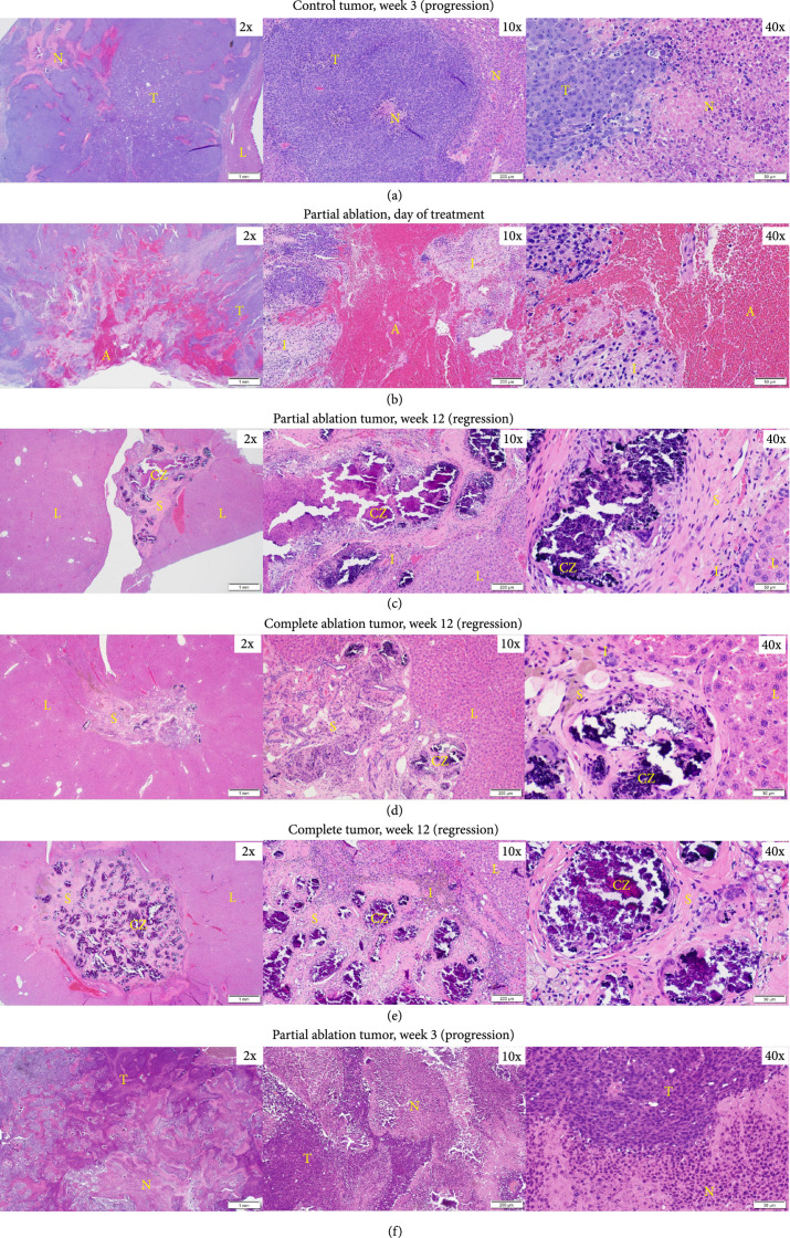
Figure 4.
Representative histology images. (a) Control HCC at 3 weeks demonstrating progression shows viable tumor (T) with prominent region of necrosis (N). (b) Partial ablation on day 0, the ablation zone (A) is comprised mainly of extravasated red blood cells and fibrin, and is surrounded by regions of inflammatory cells (I) and regions of viable tumor (T). (c) Partial ablation at 12 weeks demonstrating tumor bed completely replaced by scar tissue (S) and scattered areas of dystrophic calcification (CZ) within the ablated region with a thin rim of inflammatory cells (I) separating the ablated zone from normal liver (L) and no evidence of viable tumor. (d) Complete ablation at 3 months demonstrating tumor regression and replacement by scar tissue (S) with scattered dystrophic calcification (CZ) and a thin rim of inflammatory cells (I). No evidence of viable residual tumor in the surrounding normal liver (L). (e) Control tumor at 3 months demonstrating complete tumor regression, scattered inflammatory cells (I), surrounding scar tissue (S), and dystrophic calcification (CZ) at the original tumor site. No evidence of viable residual tumor in the surrounding normal liver (L). (f) Partial ablation at 3 weeks demonstrating tumor progression. Viable tumor (T) is observed with some regions of necrosis (N). Timepoints are measured from the histotripsy treatment time as week 0.

