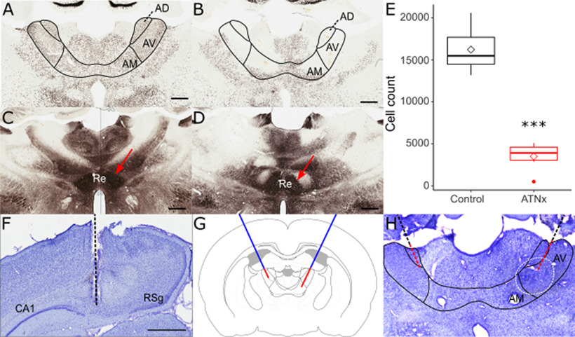
Figure 1.
NeuN-reacted coronal sections showing the status of the AV (anteroventral), AM (anteromedial) and AD (anterodorsal) thalamic nuclei in control (A) and lesion (B) animals. The nucleus reuniens (Re, arrowed), as shown using calbindin-reacted sections, was intact in both control (C) and lesion (D) animals, indicating that reuniens damage was not responsible for deficits seen in ATN-lesioned animals. E, The ATN cell count was significantly reduced in lesioned animals (ATNx) compared with controls (Control). Nissl-stained coronal sections helped to confirm electrode placement in dorsal subiculum (F), with the electrode path indicated. G, Schematic representing cannula placement (blue) and the two targets of the infusion needle (red). H, Cresyl violet-stained section indicating cannula placement, with DAB-reacted Flurogold infused to indicate spread of muscimol. Black line indicates canula placement. Red line indicates the track of the infusion needle. Dashed white line indicates the spread of the muscimol. ***p < 0.001 (Welch's two-sample t test). Scale bar, 800 mm. RSg, retrosplenial cortex.

