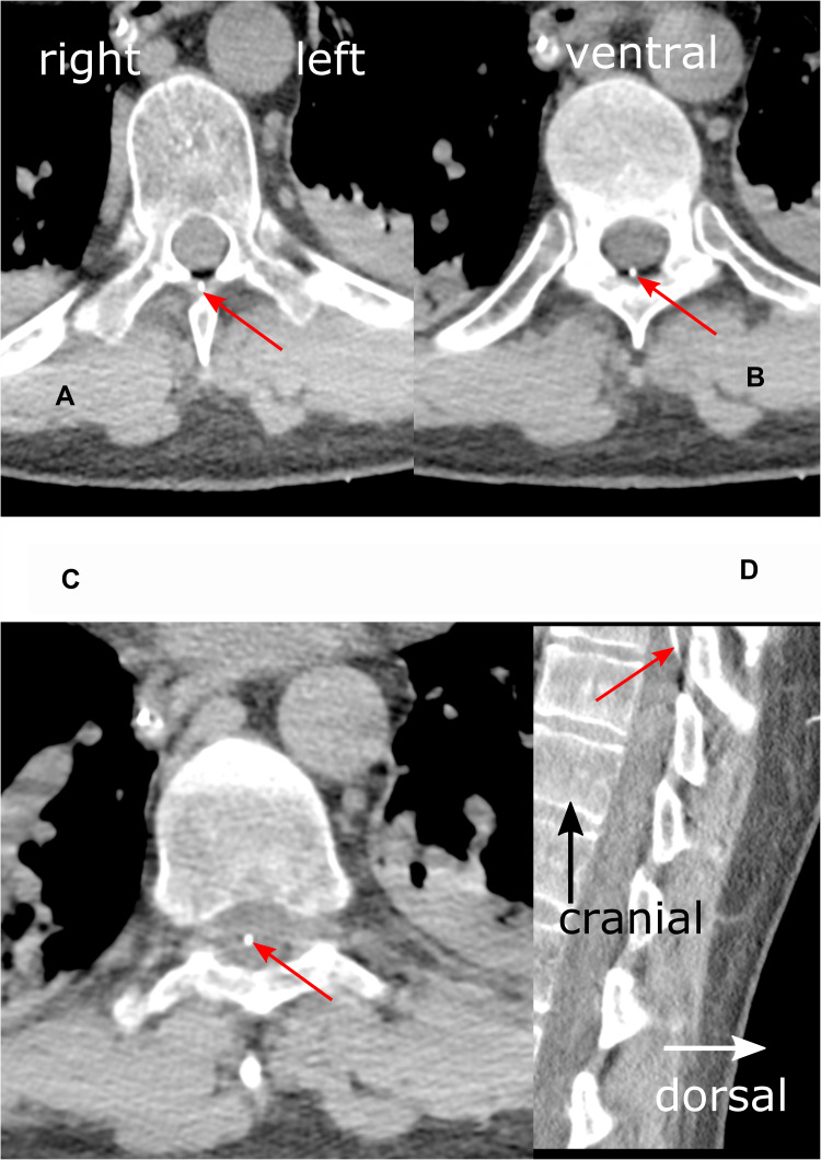
Figure 2.
Detail enlargements of axial (A–C) and sagittal (D) thoracic CT scans in a soft tissue window at level Th9. Red arrows indicate epidural catheter (white dot). (A) (left upper panel): The catheter is shown passing the ligamentum flavum ventral to the spinous process. (B) (right upper panel): Imaging of the catheter in the epidural space. (C) (left lower panel): Catheter position almost in the middle of the dural sac, suggesting a position inside the spinal cord. (D) (right lower panel): Entry of the epidural catheter into the dural sac. The terminal 4 centimeters of the catheter are not shown. Additional findings: consolidations (atelectasis) of the posterior lower lobes, nasogastric tube.

